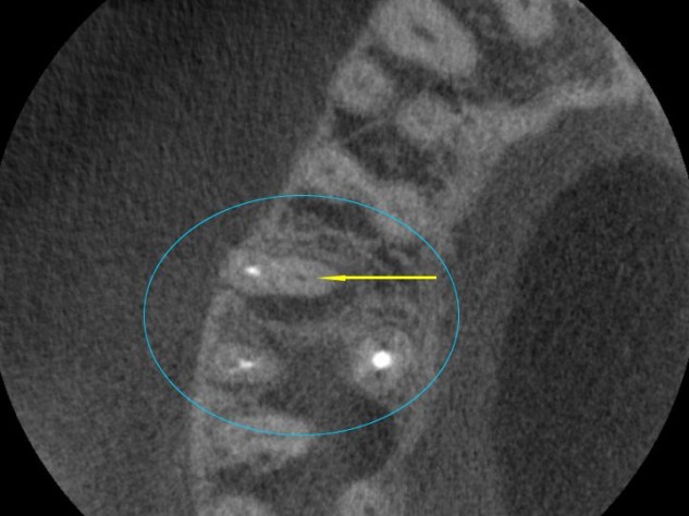
Technology » This is an axial CBCT “slice” looking at the upper right teeth. The view is as if we were standing on the bottom teeth, looking up at the top teeth, and this is a cross – section (think of looking straight down on a cut tree stump). We are looking at a previous root canal therapy that didn’t work and requires retreatment to save the tooth. The white dots are the root canal fillings. The black dot is a canal that was not treated the first time (so it is still full of bacteria) and prevented this incomplete root canal treatment from working. With CBCT, we can see this view for the first time. This offers a significant improvement in our ability diagnose and correct the problem of “missed canals” on retreatments. This view is beneficial for non-retreatment cases as well, to know how many canals are in a tooth, and their location prior to initiating root canal therapy. Teeth have a variable number of canals and all of them need to be treated for the root canal therapy to be successful.
Cone Beam Computed Tomography
