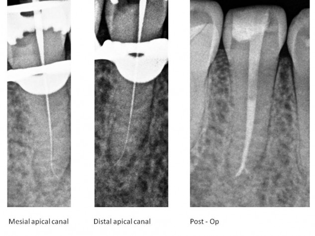
| By admin | | Comments Off on Another with more than one from yesterday. Note the mid-root and associated filled on the post-op.
While he was here, we got a 9-year on a I completed in 2011. His dentist referred after separating an instrument in the mesial root. The was removed and healing looks great on recall. Fortunately for the patient, we placed an immediate.
Sometime after we completed #19, his dentist did a on #20 and this tooth had significant decay because material had been left in place for years. Now he is set up for his permanent crowns and has scheduled with his dentist to get them done
Another #premolar with more than one #POE from yesterday. Note the mid-root #radiolucency and associated #lateralcanal filled on the post-op.
While he was here, we got a 9-year #endodonticrecall on a #rootcanalretreatment I completed in 2011. His dentist referred after separating an instrument in the mesial root. The #separatedinstrument was removed and healing looks great on recall. Fortunately for the patient, we placed an immediate #buildup.
Sometime after we completed #19, his dentist did a #pulpectomy on #20 and this tooth had significant decay because #temporaryfilling material had been left in place for years. Now he is set up for his permanent crowns and has scheduled with his dentist to get them done.
#dentistry #endodontics #endodontist
| By admin | | Comments Off on Surprise, surprise! Although I can’t see what #28 looked like before the initial RCT was done, I imagine it looked a lot like #29. This is an example of a , which is where one canal splits into two or more smaller canals. Radiographically this presents as a canal that disappears apically because the smaller canals may not be visible. So, any tooth that has this radiographic presentation should be inspected for multiple canals, even if it is a tooth that is statistically unlikely to have multiple canals like a lower premolar
Surprise, surprise! Although I can’t see what #28 looked like before the initial RCT was done, I imagine it looked a lot like #29. This is an example of a #fastbreak , which is where one canal splits into two or more smaller canals. Radiographically this presents as a canal that disappears apically because the smaller canals may not be visible. So, any tooth that has this radiographic presentation should be inspected for multiple canals, even if it is a tooth that is statistically unlikely to have multiple canals like a lower premolar. #endodontics #rootcanal #rootcanaltreatment #dentistry #endodonticretreatment
| By admin | | Comments Off on I would like to thank @sonendoinc for personally delivering sodium hypochlorite to the office so we could continue to treat emergency patients without a disruption in quality of care. This is a company that truly cares about their customers and their community
I would like to thank @sonendoinc for personally delivering sodium hypochlorite to the office so we could continue to treat emergency patients without a disruption in quality of care. This is a company that truly cares about their customers and their community.
#dentistry #gentlewave #endodontics #rootcanal
| By admin | | Comments Off on It’s nice to have good neighbors in this uncertain time.
I feel lucky to be in a community where the general dentists and specialists work together to collectively raise the bar for patient care.
The Endodontist community here is particularly special and a lot of us are good friends.
Dr. Bjorn Jonsson is a talented Endodontist in the adjacent city. Prior to the lockdown, we got together in my backyard shaping room and shaped his new board. I would say the skill and attention to detail from dentistry caries over well. The surfboard came out just as good as this lower molar RCT he treated, with a mesial and distal and.
Looking forward to this pandemic being over and life being back to normal
It’s nice to have good neighbors in this uncertain time.
I feel lucky to be in a community where the general dentists and specialists work together to collectively raise the bar for patient care.
The Endodontist community here is particularly special and a lot of us are good friends.
Dr. Bjorn Jonsson is a talented Endodontist in the adjacent city. Prior to the #coronavirus lockdown, we got together in my backyard shaping room and shaped his new board. I would say the skill and attention to detail from dentistry caries over well. The surfboard came out just as good as this lower molar RCT he treated, with a mesial #isthmus and distal #lateralcanal and #dilaceration.
Looking forward to this pandemic being over and life being back to normal.
#dentistry #dentist #endodontist #endodontics #gentlewave #worthsaving #orangecounty
| By admin | | Comments Off on Friends, if you are having a hard time finding during this @bigmachine_distillery has stepped up and is making it. You can order at bigmachinevodka.com
Big thanks to all the people and companies who are helping out during this time.
Friends, if you are having a hard time finding #handsanitizer during this #pandemic @bigmachine_distillery has stepped up and is making it. You can order at bigmachinevodka.com
Big thanks to all the people and companies who are helping out during this time.
| By admin | | Comments Off on In order to help flatten the curve for Covid-19, we have only been going in to treat true dental emergencies. The quarantine has left me with a lot of extra time for my side hustle. I started shaping surfboards in 1988 and worked through college and dental school. I still shape a few boards per week and am lucky enough to make boards for some of the best surfers in the world. In fact, Albee Layer recently won Surfer Poll awards three years in a row for the best surfing in the world each year, on boards I made in my little backyard quarantine shed. @infinity_surf @surfer_magazine
In order to help flatten the curve for Covid-19, we have only been going in to treat true dental emergencies. The quarantine has left me with a lot of extra time for my side hustle. I started shaping surfboards in 1988 and worked through college and dental school. I still shape a few boards per week and am lucky enough to make boards for some of the best surfers in the world. In fact, Albee Layer recently won Surfer Poll awards three years in a row for the best surfing in the world each year, on boards I made in my little backyard quarantine shed. @infinity_surf @surfer_magazine
| By admin | | Comments Off on I treated three cases in 2018 with persisting tapping sensitivity after conventional RCT (to my knowledge). All three resolved following going back in using gentlewave, and all three had furcal lateral canals. I recognize this is a very small and insignificant sample size. Since I started using gentlewave 14 months ago, I haven’t had any of those persisting tapping sensitivity cases. None. 🤞 Here is a recent minimally invasive gentlewave case where I’m happy to see that small furcal lateral
I treated three cases in 2018 with persisting tapping sensitivity after conventional RCT (to my knowledge). All three resolved following going back in using gentlewave, and all three had furcal lateral canals. I recognize this is a very small and insignificant sample size. Since I started using gentlewave 14 months ago, I haven’t had any of those persisting tapping sensitivity cases. None. 🤞 Here is a recent minimally invasive gentlewave case where I’m happy to see that small furcal lateral. –
#endodontics #dentistry #gentlewave #rootcanaltreatment #conservativedentistry #lateralcanal #dental #saveteeth
| By admin | | Comments Off on Three year recall from today. This patient initially came in specifically for selective retreatment of the mesial root. Apparently her sister is a dentist in Europe who practices conservative dentistry and understands that removing excess tooth structure is never a good thing. I like the conservative shapes of the original RCT, but cleaning out the canals is a problem with small shapes, and ultimately this case did not work for her. Today, I believe it is possible to thoroughly clean these shapes with . I retreated this tooth using relatively conservative shapes, a V-Taper 30, just big enough to. Excellent healing is seen at the . No unnecessary harm done to the distal, specifically at the patients request
Three year recall from today. This patient initially came in specifically for selective retreatment of the mesial root. Apparently her sister is a dentist in Europe who practices conservative dentistry and understands that removing excess tooth structure is never a good thing. I like the conservative shapes of the original RCT, but cleaning out the canals is a problem with small shapes, and ultimately this case did not work for her. Today, I believe it is possible to thoroughly clean these shapes with #gentlewave . I retreated this tooth using relatively conservative shapes, a V-Taper 30, just big enough to #endovac. Excellent healing is seen at the #endodonticrecall . No unnecessary harm done to the distal, specifically at the patients request. .
#endodontics #rootcanal #rootcanaltreatment #rootcanalretreatment #worthsaving #endoworks #savedanother
| By admin | | Comments Off on Finish from today with some good teaching points. .
Large, well defined corticated radiolucency extending into the sinus over the palatal root of #3. .
I’ve seen a lot of teeth like this taken out so the “lesion can be removed”, taken out because “cysts don’t heal”, taken out because “the prognosis of a tooth with a big lesion is poor”, etc… .
Guess what, this young lady, like most people wanted to save her tooth, and she was referred to me from a few cities away to save it. .
Even in the age of , I treated this tooth in three visits over six months and didn’t obturate until I could confirm healing. By doing this, we avoided extraction and avoided an unnecessary palatal surgery. .
I find that when done right and with enough effort, almost every case will heal, weather the lesion is big or looks like a cyst. Just check my feed or a lot of other feeds
Finish from today with some good teaching points. .
Large, well defined corticated radiolucency extending into the sinus over the palatal root of #3. .
I’ve seen a lot of teeth like this taken out so the “lesion can be removed”, taken out because “cysts don’t heal”, taken out because “the prognosis of a tooth with a big lesion is poor”, etc… .
Guess what, this young lady, like most people wanted to save her tooth, and she was referred to me from a few cities away to save it. .
Even in the age of #gentlewave , I treated this tooth in three visits over six months and didn’t obturate until I could confirm healing. By doing this, we avoided extraction and avoided an unnecessary palatal surgery. .
I find that when done right and with enough effort, almost every case will heal, weather the lesion is big or looks like a cyst. Just check my feed or a lot of other #endodontist feeds. .
#worthsaving #endoworks #rootcanaltherapy #dentistry #endodontics #nstep #endodonticrecall
| By admin | | Comments Off on This patient came in today to evaluate #14 for retreatment. .
While she was here I was able to get a 3 year recall on #15 vital pulp therapy. The pulp is vital, healthy and asymptomatic. We got to save this patient some tooth structure and money
This patient came in today to evaluate #14 for retreatment. .
While she was here I was able to get a 3 year recall on #15 vital pulp therapy. The pulp is vital, healthy and asymptomatic. We got to save this patient some tooth structure and money. .
#endodontics #vitalpulptherapy #mta #partialpulpotomy #rootcanal #dentistry #dentalpulp #pulpcap #rootcanalspecialist #stayinalive #endodonticrecall
| By admin | | Comments Off on Nice 1.5 year recall on a Nec/CAA lower incisor with a ribbon shaped canal / fin in the apical half, and a J-shaped radiolucency. Treated using and , obturated after confirming radiographic healing. Healing complete at 1.5 year
Nice 1.5 year recall on a Nec/CAA lower incisor with a ribbon shaped canal / fin in the apical half, and a J-shaped radiolucency. Treated using #endovac and #calciumhydroxide , obturated after confirming radiographic healing. Healing complete at 1.5 year #endodonticrecall –
#endoworks #worthsaving #rootcanal #endodontics #rootcanalanatomy #endodontist
| By admin | | Comments Off on Exciting 1 year recall on a challenging case, which happens to be my first case. –
This patient presented with an isolated periodontal probing to the apex at the MP of this fused mesio-palatal root, on a tooth that had RCT years before. –
These fused roots often have missed canals / anatomy in the fused part of the root, which is often difficult to locate and treat, making retreatment particularly challenging. I spent the first visit troughing this area, looking for the MB2, which would be responsible for this specific lesion. It was not found. The previous treatment had also ledged / blocked both the mb and db canals. These canals were not recovered either. –
My Gentlewave was delivered between visits so I used it at the second visit. Time was spent searching for the mb2 and trying to negotiate the mb and db canals with no luck. After running the GW I obturated with gutta percha and pulp canal sealer with warm vertical compaction. The MB2 was seen on the post-op CBCT splitting off from within the palatal canal. To my surprise, sealer also filled the DB canal to the apex. –
The one year recall shows excellent healing. Periodontal probing is 3 mm
Exciting 1 year recall on a challenging case, which happens to be my first #gentlewave case. –
This patient presented with an isolated periodontal probing to the apex at the MP of this fused mesio-palatal root, on a tooth that had RCT years before. –
These fused roots often have missed canals / anatomy in the fused part of the root, which is often difficult to locate and treat, making retreatment particularly challenging. I spent the first visit troughing this area, looking for the MB2, which would be responsible for this specific lesion. It was not found. The previous treatment had also ledged / blocked both the mb and db canals. These canals were not recovered either. –
My Gentlewave was delivered between visits so I used it at the second visit. Time was spent searching for the mb2 and trying to negotiate the mb and db canals with no luck. After running the GW I obturated with gutta percha and pulp canal sealer with warm vertical compaction. The MB2 was seen on the post-op CBCT splitting off from within the palatal canal. To my surprise, sealer also filled the DB canal to the apex. –
The one year recall shows excellent healing. Periodontal probing is 3 mm. –
#endodontics #rootcanaltreatment #rootcanal #endodonticretreatment #endodontist #endodonticrecall #notaverticalrootfracture #mb2 #dentistry #rootcanalanatomy
| By admin | | Comments Off on Previous RCT ✅
Deep, narrow probing ✅
J-shaped radiolucency ✅
Vertical root fracture 🚫
–
Another save, thanks to this patients for referring. The first step in these cases is to look to see if a fracture is present, not extracting after guessing one is present. After accessing and confirming no fracture is present, then this becomes a very predictable retreatment case. Bottom images show good healing at 8 month recall
Previous RCT ✅
Deep, narrow probing ✅
J-shaped radiolucency ✅
Vertical root fracture 🚫
–
Another save, thanks to this patients #dentist for referring. The first step in these cases is to look to see if a fracture is present, not extracting after guessing one is present. After accessing and confirming no fracture is present, then this becomes a very predictable retreatment case. Bottom images show good healing at 8 month recall-
#endodonticretreatment #rootcanaltreatment #rootcanal #rootcanalspecialist #endodontics #endodontist #endodonticrecall #verticalrootfracture #dentalimplant #dentalhygiene #periodontics #endoperiolesion #itsnotcracked #stopassumingitscracked
| By admin | | Comments Off on PA: Just another basic premolar .
CBCT: Hess anatomy .
–
Fortunately this type of anatomy is now predictable to treat with the incorporation of advanced technology in our treatment protocol
PA: Just another basic premolar .
CBCT: Hess anatomy .
–
Fortunately this type of anatomy is now predictable to treat with the incorporation of advanced technology in our treatment protocol . #gentlewave #cbct #dentaloperatingmicroscope –
#endodontist #endodontics #dentistry #rootcanaltreatment #rootcanal #endodonticanatomy #hessanatomy
| By admin | | Comments Off on My emergency patient this morning happened to be the patient from the #3 case I posted last week .
–
We took a for tooth #3, which shows complete healing, even with this complex .
–
It turns out her #18 had pretty wild as well
My emergency patient this morning happened to be the patient from the #3 #endodance case I posted last week .
–
We took a #endodonticrecall #cbct for tooth #3, which shows complete healing, even with this complex #endodonticanatomy .
–
It turns out her #18 had pretty wild #apicalanatomy as well. –
#endodontics #endodontia #endodoncia #dentistry #rootcanal #rootcanaltherapy #gentlewave
| By admin | | Comments Off on One of the most rewarding case types, for a number of reasons.
This patient had failing RCTs on #15 and 16 four years after being treated by an Endodontist. She returned to that Endodontist, who removed the crown on 15 and made a large access in the crown on 16, and still was unable to find the canals. They put in a good effort to find the missed canal in 15, but were looking in the wrong place. Fortunately the roots are fused so no perforation was created. At this point the towel was thrown in and patient was advised to extract both teeth.
She was very upset at the idea of losing these teeth, even with one of them being a third molar. So she found me for a second opinion.
I treated these cases in multiple visits to make sure it they were working prior to finishing. Sometimes just a little more effort is all the difference. These are cases where anybody’s “best hour” isn’t good enough, these require time and effort.
Slide 1: Pre-op images. Note area of deep troughing in 15, and untreated DB canals in both teeth.
Slide 2: located troughed area, filled with MTA, placed composite over MTA, located missed DB canal.
Slide 3: previous (large) access in 16, located untreated DB canal. Removed all gutta percha in both teeth, located, shaped and all canals prior to placing
Slide 4: Visit 3. After a calcium hydroxide change at visit 2. Images were taken that confirmed healing (Images on right). The cases were obturated at this visit, only after healing was confirmed radiographically. This eliminates 🤞. Slide 5. One year recall on right shows excellent healing. This patient was extremely happy we took the time and effort to save her teeth, even her wisdom tooth.
Slide 6: a quote that Gary Carr sent me that I always think about
One of the most rewarding case types, for a number of reasons.
This patient had failing RCTs on #15 and 16 four years after being treated by an Endodontist. She returned to that Endodontist, who removed the crown on 15 and made a large access in the crown on 16, and still was unable to find the canals. They put in a good effort to find the missed canal in 15, but were looking in the wrong place. Fortunately the roots are fused so no perforation was created. At this point the towel was thrown in and patient was advised to extract both teeth.
She was very upset at the idea of losing these teeth, even with one of them being a third molar. So she found me for a second opinion.
I treated these cases in multiple visits to make sure it they were working prior to finishing. Sometimes just a little more effort is all the difference. These are cases where anybody’s “best hour” isn’t good enough, these require time and effort.
Slide 1: Pre-op images. Note area of deep troughing in 15, and untreated DB canals in both teeth.
Slide 2: located troughed area, filled with MTA, placed composite over MTA, located missed DB canal.
Slide 3: previous (large) access in 16, located untreated DB canal. Removed all gutta percha in both teeth, located, shaped and #endovac all canals prior to placing #calciumhydroxide
Slide 4: Visit 3. After a calcium hydroxide change at visit 2. Images were taken that confirmed healing (Images on right). The cases were obturated at this visit, only after healing was confirmed radiographically. This eliminates 🤞. Slide 5. One year recall on right shows excellent healing. This patient was extremely happy we took the time and effort to save her teeth, even her wisdom tooth.
Slide 6: a quote that Gary Carr sent me that I always think about.
#endodontics #endodontia #endodoncia #dentistry #dentalcases #rootcanal #rootcanaltreatment #rootcanalretreatment #missedcanal #nstep #endodonticrecall
| By admin | | Comments Off on When you don’t want to go to the dentist…but your brothers are there to lend their support. –
@jaredsislinphotography @teamboehne❤️
When you don’t want to go to the dentist…but your brothers are there to lend their support. –
@jaredsislinphotography @teamboehne #myfamily❤️
| By admin | | Comments Off on Not to be outdone by the from the in my last post. My gave me this one this morning. I’ve got to say, the technology made this one 100% easier, more conservative, and I believe more predictable
Not to be outdone by the #apicalanatomy from the #endodance in my last post. My #gentlewave gave me this one this morning. I’ve got to say, the technology made this one 100% easier, more conservative, and I believe more predictable. –
#endodontics #endodonticirrigation #endodontist #rootcanal #rootcanaltherapy #dentistry #apicaldilaceration #apicalbifurcation #apicaltrifurcation #apicaldelta
| By admin | | Comments Off on A case from a few years ago, before I was using and . In this case, the preoperative showed complex anatomy with a lateral canal in the palatial root and multiple in the . The anatomy was treated with apical negative pressure irrigation as well as sonic activated irrigation, and the “endo dance”. Obturated using with
A case from a few years ago, before I was using #gentlewave and #bcsealer . In this case, the preoperative #cbct showed complex anatomy with a lateral canal in the palatial root and multiple #portalsofexit in the #mbroot . The anatomy was treated with #endovac apical negative pressure irrigation as well as #endoactivator sonic activated irrigation, and the “endo dance”. Obturated using #warmverticalcompaction with #pulpcanalsealer. –
#endodontics #endodontia #endodoncia #dental #dentistry #dentalanatomy #rootcanal #rootcanaltherapy
| By admin | | Comments Off on I saw my friend @karenpotterdds post about a molar HRF earlier today, then this patient walked in the door after. As far as I can recall it’s the first HRF on a molar I’ve seen. What are the odds?
–
This patient came in for #4, which has a split tooth that happened when she was eating the other day. This tooth will be extracted. –
The PA and CBCT also show a on the P root of #3. Pt hasn’t had an opposing tooth for a decade. She denies a history of trauma. This tooth is asymptomatic and has a healthy vital pulp. The fracture pattern is favorable, remaining subosseous, preventing infection. No treatment is recommended for #3
I saw my friend @karenpotterdds post about a molar HRF earlier today, then this patient walked in the door after. As far as I can recall it’s the first HRF on a molar I’ve seen. What are the odds?
–
This patient came in for #4, which has a split tooth that happened when she was eating the other day. This tooth will be extracted. –
The PA and CBCT also show a #HRF on the P root of #3. Pt hasn’t had an opposing tooth for a decade. She denies a history of trauma. This tooth is asymptomatic and has a healthy vital pulp. The fracture pattern is favorable, remaining subosseous, preventing infection. No treatment is recommended for #3. –
#endodontics #dentistry #horizontalrootfracture #splittooth #crackedtooth #dentalimplant #endodontist
| By admin | | Comments Off on Apical bifurcations du jour. Buildup placed with in palatal root. Initial crown margin prepared. I like to make life easy for the referring doc and the case predictable for the patient
Apical bifurcations du jour. Buildup placed with #fiberpost in palatal root. Initial crown margin prepared. I like to make life easy for the referring doc and the case predictable for the patient. –
#dentistry #endodontics #endodontist #apicalbifurcation #rootcanal #rootcanaltherapy
| By admin | | Comments Off on Teaching my boy a couple important lessons like trusting Dad, facing your fears, making friends, respecting nature, but most importantly learning about class III malocclusion
[dsgnwrks_instagram_embed src=”https://www.instagram.com/p/B4_nrXeJztT/” type=”video”]
Teaching my boy a couple important lessons like trusting Dad, facing your fears, making friends, respecting nature, but most importantly learning about class III malocclusion. #prognathism #thatsabigeel
| By admin | | Comments Off on Extensive carious lesion.
Pretty much the only remaining tooth structure is the lingual surface / cingulum.
Pt understands the guarded long-term prognosis but would prefer to save this tooth for as long as possible.
Caries directed access. No healthy tooth structure was removed for convenience form or an “ideal”endodontic access. Cingulum and lingual surface preserved.
Perfect case for. It is possible to thoroughly irrigate to the apex without hypochlorite touching the gingiva, which would cause bleeding and compromise bonding.
A rare case where a create a post space larger than the existing V-taper shape, to fit a slightly larger.
Tooth removed from occlusion. A crown is contraindicated because it would remove the only remaining tooth structure
Extensive carious lesion.
Pretty much the only remaining tooth structure is the lingual surface / cingulum.
Pt understands the guarded long-term prognosis but would prefer to save this tooth for as long as possible.
Caries directed access. No healthy tooth structure was removed for convenience form or an “ideal”endodontic access. Cingulum and lingual surface preserved.
Perfect case for #endovac. It is possible to thoroughly irrigate to the apex without hypochlorite touching the gingiva, which would cause bleeding and compromise bonding.
A rare case where a create a post space larger than the existing V-taper shape, to fit a slightly larger #fiberpost.
Tooth removed from occlusion. A crown is contraindicated because it would remove the only remaining tooth structure.
#endodontics #herodontics #caries #errrairbubblebehindthepost #crowding
| By admin | | Comments Off on A little friend showed up in front of our office Monday afternoon! Video: @paytonlandaas
[dsgnwrks_instagram_embed src=”https://www.instagram.com/p/B4268HxJ5db/” type=”video”]
A little friend showed up in front of our office Monday afternoon! Video: @paytonlandaas
| By admin | | Comments Off on 10-month healing on a case with a in the apical third of the root. This case initially probed to the apex and now probes 3 mm. –
The file was removed by bypassing with pre-curved stainless steel k-files, then it was teased loose and removed with
–
Another save by the . How many of these teeth never make it to the Endodontist and just get extracted? This patient is very grateful that her dentist referred her to us
10-month healing on a case with a #separatedinstrument in the apical third of the root. This case initially probed to the apex and now probes 3 mm. –
The file was removed by bypassing with pre-curved stainless steel k-files, then it was teased loose and removed with #endovac
–
Another save by the #Endodontist . How many of these teeth never make it to the Endodontist and just get extracted? This patient is very grateful that her dentist referred her to us. #endodontistssaveteeth –
#endodontics #dentistry #dentalhygiene #oralsurgery #periodontics #rootcanal #endodontics #verticalrootfracture #dentalimplant #saveteeth #separatedfile #endodonticrecall
| By admin | | Comments Off on #14. Another amazing case by Dr. Chu. Note that the complex anatomy, including the apical bifurcation in the MB root, was cleaned and obturated with dentin-preserving conservative root shapes. This case also illustrates that endodontic access cavity designs can still be very conservative when using @wcendo
#14. Another amazing case by Dr. Chu. Note that the complex anatomy, including the apical bifurcation in the MB root, was cleaned and obturated with dentin-preserving conservative root shapes. This case also illustrates that endodontic access cavity designs can still be very conservative when using #gentlewave –
#endodontist #endodontics #endodontia #endodoncia #sonendo #rootcanal #rootcanalanatony #endodonticaccess #biomimeticdentistry #apicalbifurcation @wcendo
| By admin | | Comments Off on Kind of a long story…
.
Slide 1: #18 Probes to apex. Nec/chronic apical abscess draining through gingival sulcus. Just based on the lateral boneloss how many oral surgeons would take this tooth out? What a mistake and disservice. –
Slide 2: immediate post-op PA. No cracks seen through the microscope. Used conservative root preps to preserve tooth structure and minimize the chance of future root fracture. –
Slide 3: healed on PA and CBCT at 6 mo recall. I already confirmed the probing healed at the second visit, prior to finishing. –
Slide 4: Pt returns a year later, it probes to the apex again. CBCT shows the extent of boneloss along the length of the mesial root. Narrow isolated probing. How many people take it out now? How many people kicked themselves for not taking it out before? What about those conservative root shapes minimizing the chance of root fractures?
–
Slide 5: But wait! The old sinus tracing trick traces to the apex of #19. #19 became necrotic and the abscess of #19 was draining through the sulcus of #18. –
Slide 6: both teeth completely healed and probing on #18 returned to 2 mm. –
This patient was so happy that we saved his teeth. We take the time to think these things through and make the right treatment decisions. In the wrong hands, both of these teeth could have been extracted. No person prefers that. Endodontists save teeth
Kind of a long story…
.
Slide 1: #18 Probes to apex. Nec/chronic apical abscess draining through gingival sulcus. Just based on the lateral boneloss how many oral surgeons would take this tooth out? What a mistake and disservice. –
Slide 2: immediate post-op PA. No cracks seen through the microscope. Used conservative #trushape root preps to preserve tooth structure and minimize the chance of future root fracture. –
Slide 3: healed on PA and CBCT at 6 mo recall. I already confirmed the probing healed at the second visit, prior to finishing. –
Slide 4: Pt returns a year later, it probes to the apex again. CBCT shows the extent of boneloss along the length of the mesial root. Narrow isolated probing. How many people take it out now? How many people kicked themselves for not taking it out before? What about those conservative root shapes minimizing the chance of root fractures?
–
Slide 5: But wait! The old #guttapercha sinus tracing trick traces to the apex of #19. #19 became necrotic and the abscess of #19 was draining through the sulcus of #18. –
Slide 6: both teeth completely healed and probing on #18 returned to 2 mm. –
This patient was so happy that we saved his teeth. We take the time to think these things through and make the right treatment decisions. In the wrong hands, both of these teeth could have been extracted. No person prefers that. Endodontists save teeth. –
#endodontists #endodontics #endodontia #endodoncia #dentistry #periodontics #oralsurgery #dentalimplants #rootcanaltherapy #rootcanal #verticalrootfracture #endodonticrecall #dentalcases #dentalhygiene
| By admin | | Comments Off on This patient was referred because #19 had an abscess and an isolated deep probing to the apex of the mesial root. Fortunately her dentist didn’t extract #19, but referred her to me to see if the root was cracked. .
It turns out #19 was fine. The actual problem was #20 had pulpal necrosis with a chronic apical abscess draining through the sulcus of #19
This patient was referred because #19 had an abscess and an isolated deep probing to the apex of the mesial root. Fortunately her dentist didn’t extract #19, but referred her to me to see if the root was cracked. .
It turns out #19 was fine. The actual problem was #20 had pulpal necrosis with a chronic apical abscess draining through the sulcus of #19.
.
#endodontics #endodontist #endodontia #endodoncia #rootcanal #oralsurgery #verticalrootfracture #periodontics #dentistry #dentalhygiene #dentalimplant #saveteeth #worthsaving
| By admin | | Comments Off on The perfect scenario. This is a complex C-shaped root canal system with a lot of anatomy that files wouldn’t touch. Irrigation is probably more important in this case type than most. Length control is critical because the apical construction is far from the radiographic apex. It is in fact divergent if you are only using a PA for length determination, and it happens to be right over the Inferior alveolar nerve. This makes for a risky situation for positive pressure irrigation and obturation. In this situation negative pressure irrigation is the way to go. In the past, I would have treated this case with endovac, but would have created apical shape to fit the microcannula, risking loss of the apical construction, potentially leaving a more divergent apex over the IAN. In this case I was able to keep the files far short of the apex and allowed the GW to thoroughly clean out the entire system, then comfortably obturated to the natural apical construction
The perfect #gentlewave scenario. This is a complex C-shaped root canal system with a lot of anatomy that files wouldn’t touch. Irrigation is probably more important in this case type than most. Length control is critical because the apical construction is far from the radiographic apex. It is in fact divergent if you are only using a PA for length determination, and it happens to be right over the Inferior alveolar nerve. This makes for a risky situation for positive pressure irrigation and obturation. In this situation negative pressure irrigation is the way to go. In the past, I would have treated this case with endovac, but would have created apical shape to fit the microcannula, risking loss of the apical construction, potentially leaving a more divergent apex over the IAN. In this case I was able to keep the files far short of the apex and allowed the GW to thoroughly clean out the entire system, then comfortably obturated to the natural apical construction. –
#endodontics #endodontist #cshapedmolar #rootcanal #rootcanalanatomy #dentistry
| By admin | | Comments Off on 8 month recall of a case. Nec/CAA. J-shaped radiolucency, proved to the apex. Healed at 8 months. Another example that shows how often there’s aren’t vertical root fractures
8 month recall of a #gentlewave case. Nec/CAA. J-shaped radiolucency, proved to the apex. Healed at 8 months. Another example that shows how often there’s aren’t vertical root fractures. –
#endodontics #endodontist #verticalrootfracture #dentalimplants #periodontics #endodonticrecall #jshapedlesion #rootcanaltreatment #rootcanal #dentistry
| By admin | | Comments Off on Dr. Chu coming through with a 2:1:2 today. –
Also check out the #30 he did at his other office @valleygreenendodontics yesterday @wcendo
Dr. Chu coming through with a 2:1:2 today. –
Also check out the #30 he did at his other office @valleygreenendodontics yesterday
–
#rootcanalspecialist #endodontist #endodontics #rootcanalanatomy #apicaldilaceration #molar #premolar #dentistry #drchucomingthru #iwashisbenchinstructor @wcendo
| By admin | | Comments Off on There are still a couple spots left in the @smileonu event in Huntington Beach. All proceeds go to this really great organization and the entry fee is a tax deduction.
There are still a couple spots left in the @smileonu_ #surfforcharity event in Huntington Beach. All proceeds go to this really great organization and the entry fee is a tax deduction.
| By admin | | Comments Off on You know that was a fun case based on the pre-op PA. A lot of time spent carefully and thoughtfully negotiating and shaping these multi-plane curvatures
You know that was a fun case based on the pre-op PA. A lot of time spent carefully and thoughtfully negotiating and shaping these multi-plane curvatures. –
#endodontics #endodontia #endodoncia #rootcanal #rootcanaltherapy #rootcanalanatomy #dentistry #uclasod #radixentemolaris
| By admin | | Comments Off on A couple years before I got on the bandwagon
A couple years before I got on the #gentlewave bandwagon. –
#hurricanemarie #thewedge #endodontist #notsogentlewave #stillmadeittowork #dentistry #rootcanaltreatment
| By admin | | Comments Off on The likelihood of this case type failing and this patient extracting this tooth in the future isn’t from recurring infection or what we think of as a true Endodontic failure. It’s from tooth fracture, specifically cervical snap off; a restorative failure. These tend to happen down the road and the small details for long term success are often overlooked. –
If the only intercuspal coronal tooth structure remaining after interproximal caries removal is directly over the root canal, you can preserve this tooth structure by changing “ideal access” to a caries-directed access. Convenience form has no role here. –
A fiber post is used to help retain the restoration and to some degree minimize the risk for cervical snap off. A fiber post can be placed here without developing the root-cracking high stress concentration that a short off axis metal post would, due to its modulus of elasticity being comparable to dentin. –
Another tip: whenever this referring dentist has a various pulp exposure, she temporize with IRM directly over the pulp and none of the patients develop pain
The likelihood of this case type failing and this patient extracting this tooth in the future isn’t from recurring infection or what we think of as a true Endodontic failure. It’s from tooth fracture, specifically cervical snap off; a restorative failure. These tend to happen down the road and the small details for long term success are often overlooked. –
If the only intercuspal coronal tooth structure remaining after interproximal caries removal is directly over the root canal, you can preserve this tooth structure by changing “ideal access” to a caries-directed access. Convenience form has no role here. –
A fiber post is used to help retain the restoration and to some degree minimize the risk for cervical snap off. A fiber post can be placed here without developing the root-cracking high stress concentration that a short off axis metal post would, due to its modulus of elasticity being comparable to dentin. –
Another tip: whenever this referring dentist has a various pulp exposure, she temporize with IRM directly over the pulp and none of the patients develop pain. –
#endodontics #endorestorative #fiberpost #dentistry #rootcanal #endodontia #endodoncia #rootcanaltherapy #thelittlethings
| By admin | | Comments Off on If endo was a cook book recipe, this tooth would be in the garbage.
Some clinicians recommend filling to the radiographic terminus on every case. In this case the canals exited the root 3-4 mm short of the radiographic apex on the mesial root. Achieving “the look” would have meant extruding the cones into the surrounding bone, sacrificing the biology of Endodontics for the “art” of Endodontics.
The distal root has internal resorption. Imagine what a disaster creating a continuous tapering funnel for a process outcome would have been here.
In this case the and helped me to establish correct lengths. I used to clean the irregularities of this root canal system.
The look is ugly but the prognosis is excellent
If endo was a cook book recipe, this tooth would be in the garbage.
Some clinicians recommend filling to the radiographic terminus on every case. In this case the canals exited the root 3-4 mm short of the radiographic apex on the mesial root. Achieving “the look” would have meant extruding the cones into the surrounding bone, sacrificing the biology of Endodontics for the “art” of Endodontics.
The distal root has internal resorption. Imagine what a disaster creating a continuous tapering funnel for a process outcome would have been here.
In this case the #cbct and #apexlocator helped me to establish correct lengths. I used #gentlewave to clean the irregularities of this root canal system.
The look is ugly but the prognosis is excellent.
#theartofendodontics #endodontics #rootcanal #endodontist #dentistry #internalresorption #inflammatoryresorption #trustyourapexlocatoroverthepa
| By admin | | Comments Off on My favorite lecture of the year. 3rd years are officially ready for clinic #13041
My favorite lecture of the year. 3rd years are officially ready for clinic. –
#dentistry #endodontics #ucla #uclasod #endodonticcomplication #rootcanal #13041
| By admin | | Comments Off on reached the pulp of #25 causing irreversible pulpitis. With cervical tooth structure missing due to the resorptive process, all remaining cervical tooth structure should be preserved to minimize the risk of cervical snap off. This includes the cingulum, which would have been weakened in a cingulum based access, particularly in a lower incisor with a lingual branch. The complex 1:2:1 root canal system was easily treated through a conservative 1.1mm incisal access. Two cones were down packed individually because they would not fit simultaneously. –
I think I’ve posted the rubber dam clamp trick for reflecting tissue during these surgeries
#cervicalinvasiveresorption reached the pulp of #25 causing irreversible pulpitis. With cervical tooth structure missing due to the resorptive process, all remaining cervical tooth structure should be preserved to minimize the risk of cervical snap off. This includes the cingulum, which would have been weakened in a cingulum based access, particularly in a lower incisor with a lingual branch. The complex 1:2:1 root canal system was easily treated through a conservative 1.1mm incisal access. Two cones were down packed individually because they would not fit simultaneously. –
I think I’ve posted the rubber dam clamp trick for reflecting tissue during these surgeries. –
#endodontics #endodontist #dentistry #rootcanal #resorption #endodonticaccess #dental
| By admin | | Comments Off on SmileOnU is a humanitarian organization donating dental care around the world. SmileOnU is supported by volunteer dental providers. I have had the opportunity to work with this great organization and see what an impact they have.
Please join SmileOnU 1st Annual Surf for Charity 🏄♂️. There will be a surf contest -for all levels, even beginners- / surf session followed brunch. I will be there offering free surf lessons and I’m giving away a surfboard at the event – more about that on the next post. Everyone is invited! Come spend a fun day surfing in Huntington Beach while supporting a good cause!
Saturday October 12th I 🏖 Huntington Beach, Pier 1
Surfing Event: 9am-10:30am
Brunch: 11am-12:30pm
Fred’s Mexican Café & Cantina
Participants will automatically be entered to win a ‘Custom Surfboard’ 🏄♂️ Adult Entry fee $300.00
Kids 15 and under) $25.00
(brunch will be provided)
To sign-up please go to SmileOnU.org to register
@smileonu_
SmileOnU is a humanitarian organization donating dental care around the world. SmileOnU is supported by volunteer dental providers. I have had the opportunity to work with this great organization and see what an impact they have.
Please join SmileOnU 1st Annual Surf for Charity 🏄♂️. There will be a surf contest -for all levels, even beginners- / surf session followed brunch. I will be there offering free surf lessons and I’m giving away a surfboard at the event – more about that on the next post. Everyone is invited! Come spend a fun day surfing in Huntington Beach while supporting a good cause!
Saturday October 12th I 🏖 Huntington Beach, Pier 1
Surfing Event: 9am-10:30am
Brunch: 11am-12:30pm
Fred’s Mexican Café & Cantina
Participants will automatically be entered to win a ‘Custom Surfboard’ 🏄♂️ Adult Entry fee $300.00
Kids 15 and under) $25.00
(brunch will be provided)
To sign-up please go to SmileOnU.org to register
@smileonu_
| By admin | | Comments Off on “3D” disinfection and tissue dissolution is by far the most important factor in the endodontic triad. Today, I mostly use for disinfection and tissue dissolution, but we were still achieving this goal in the past. –
This case was used in Cohenca’s Disinfection of Root Canal Systems to demonstrate the effectiveness of apical negative pressure irrigation at cleaning lateral anatomy. I treated this vital case with 9 POEs in one visit using
“3D” disinfection and tissue dissolution is by far the most important factor in the endodontic triad. Today, I mostly use #gentlewave for disinfection and tissue dissolution, but we were still achieving this goal in the past. –
This case was used in Cohenca’s Disinfection of Root Canal Systems to demonstrate the effectiveness of apical negative pressure irrigation at cleaning lateral anatomy. I treated this vital case with 9 POEs in one visit using #endovac
–
#endodontics #endodontia #rootcanal #rootcanaltreatment #endodontist #dentistry #rootcanalanatomy
| By admin | | Comments Off on Beautifully managed case by Dr. Chu from this morning. Severe mid-root curve on a 29 mm premolar.
–
@wcendo
Beautifully managed case by Dr. Chu from this morning. Severe mid-root curve on a 29 mm premolar.
–
@wcendo #endodontics #endodontist #endodontia #rootcanaltreatment #rootcanal #dentistry #rootcanalanatomy #vtaper2h #gentlewave #personalspace #overlap
| By admin | | Comments Off on The goal of is to remove becteria and debris from the root canal system, without damaging the tooth in the process, and preventing new bacteria from coming in. Root canals will fail for certain reasons that are completely avoidable such as missed canals and leaking temporary restorations. The risk of other factors such as inadequate disinfection and future root fracture can be significantly minimized with the right treatment strategy. .
Anatomy of predictable Endodontics:
✅Pre-op CBCT to ensure no anatomy will be missed or shaped inappropriately ✅Conservative shapes with files preserving pericervical dentin and maintaining the s-curve
✅Thorough disinfection with ✅3D with and sealer
✅immediate coronal seal. ✅perfectly fit existing crown by an excellent restorative dentist
The goal of #rootcanaltherapy is to remove becteria and debris from the root canal system, without damaging the tooth in the process, and preventing new bacteria from coming in. Root canals will fail for certain reasons that are completely avoidable such as missed canals and leaking temporary restorations. The risk of other factors such as inadequate disinfection and future root fracture can be significantly minimized with the right treatment strategy. .
Anatomy of predictable Endodontics:
✅Pre-op CBCT to ensure no anatomy will be missed or shaped inappropriately ✅Conservative shapes with #vtaper2h files preserving pericervical dentin and maintaining the s-curve
✅Thorough disinfection with #gentlewave ✅3D #obturation with #guttapercha and #bioceramic sealer
✅immediate coronal seal. ✅perfectly fit existing crown by an excellent restorative dentist .
#endodontics #endodontia #dentistry #endodontist #rootcanal #rootcanalanatomy #apicaldilaceration #apicaldelta
| By admin | | Comments Off on Six month recall of a Nec/CAA case. This case probed to the apex on both roots, and predictable healed following .
Shaped to a V-taper 30, irrigation with
Six month recall of a Nec/CAA case. This case probed to the apex on both roots, and predictable healed following #endodontictreatment .
Shaped to a V-taper 30, irrigation with #endovac .
#saveteeth #rootcanalswork #notaverticalrootfracture #endodonticrecall #rootcanal #dentalimplant #dentistry #endodontics #endodontist
| By admin | | Comments Off on This is what happens when extreme athlete and Go Pro team rider cracks a tooth –
Reposted from @chuckpatterson @gopro –
*selfie taken by professionals in a controlled environment, don’t try this at home
This is what happens when extreme athlete and Go Pro team rider #chuckpatterson cracks a tooth. –
#extremeselfie #dentistselfie #gopro #hero4session –
Reposted from @chuckpatterson @gopro –
*selfie taken by professionals in a controlled environment, don’t try this at home
| By admin | | Comments Off on This patient came in the other day for RCT #18 due to irreversible pulpitis. This gave us a 6-year recall on tooth #19, which I treated in 2013. .
In 2013 she was referred to me to evaluate #18 for a possible vertical root fracture that the dentist suspected due to an isolated probing to the apex on the mesial root. .
My thoughts today are that this patient is lucky because many dentists would have extracted #18 thinking it had a , placed a , then later extracted #19 when they found out it was failing and placed another implant. She is so much happier to keep her natural teeth. –
Im also glad to see how much more conservative endodontic shapes have become. I likely used in 2013, and the shapes from last week are
This patient came in the other day for RCT #18 due to irreversible pulpitis. This gave us a 6-year recall on tooth #19, which I treated in 2013. .
In 2013 she was referred to me to evaluate #18 for a possible vertical root fracture that the dentist suspected due to an isolated probing to the apex on the mesial root. .
My thoughts today are that this patient is lucky because many dentists would have extracted #18 thinking it had a #verticalrootfracture , placed a #dentalimplant , then later extracted #19 when they found out it was failing and placed another implant. She is so much happier to keep her natural teeth. –
Im also glad to see how much more conservative endodontic shapes have become. I likely used #protaper in 2013, and the shapes from last week are #vtaper2h .
#endodontics #endodoncia #rootcanal #endodontist #dentistry #oralsurgery #periodontics #endodonticrecall #saveteeth
| By admin | | Comments Off on Thanks for the responses on the previous case. Here is how I treated it. .
Non-surgical retreatment was completed in four visits, over 11 months. The images are from the day of obturation, after confirming bone fill. I look forward to seeing future recalls
Thanks for the responses on the previous case. Here is how I treated it. .
Non-surgical retreatment was completed in four visits, over 11 months. The images are from the day of obturation, after confirming bone fill. I look forward to seeing future recalls. .
#endodontics #endodonticrecall #endodonticretreatment #dentistry #rootcanaltreatment #saveteeth
| By admin | | Comments Off on #7 Previous RCT, very large radiolucent finding. Displaced root? Patient was swollen and experiencing discomfort. Adjacent teeth had normal responses to cold. .
What would you do?
.
A) Retreat 7
B) RCT adjacent teeth
C) Apical surgery
D) Refer to OS for extraction / biopsy?
E) Something else? What?
#7 Previous RCT, very large radiolucent finding. Displaced root? Patient was swollen and experiencing discomfort. Adjacent teeth had normal responses to cold. .
What would you do?
.
A) Retreat 7
B) RCT adjacent teeth
C) Apical surgery
D) Refer to OS for extraction / biopsy?
E) Something else? What?
.
#endodontics
#dentistry #dentalimplants #dentaleducation #endodonticretreatment #apicalsurgery #oralsurgery
| By admin | | Comments Off on The previous case was a case I did a few years back and was written up in the Endodontics chapter of Avoiding and Treating Dental Complications – Best Practices in Dentistry. I had the privilege and honor of writing with my good friend and mentor Dr. Shane White from the . You can pick up a copy on Amazon
The previous case was a case I did a few years back and was written up in the Endodontics chapter of Avoiding and Treating Dental Complications – Best Practices in Dentistry. I had the privilege and honor of writing with my good friend and mentor Dr. Shane White from the #uclaschoolofdentistry . You can pick up a copy on Amazon. –
#endodontics #endodontia #endodoncia #dentistry #dentaleducation #dentalschool #rootcanaltreatment
| By admin | | Comments Off on Teaching case. Recent dental school grad initiates RCT #9 and perforates, temporizes, then refers to us. The two critical mistakes were (1) assuming that this tooth was easy: not appreciating the receded canal, and (2) not understanding the proper angle of access. –
A) Initial presentation. Note how the access angle guaranteed missing the canal and perforation.
B) Cavit
C) Access angle leading to perforation
D) White MTA
E) Perforation repair
F) Note the proper angle used to locate canal
G) Canal obturated
H) Post-op
I) Healing at the one year
Teaching case. Recent dental school grad initiates RCT #9 and perforates, temporizes, then refers to us. The two critical mistakes were (1) assuming that this tooth was easy: not appreciating the receded canal, and (2) not understanding the proper angle of access. –
A) Initial presentation. Note how the access angle guaranteed missing the canal and perforation.
B) Cavit
C) Access angle leading to perforation
D) White MTA #perforationrepair
E) Perforation repair
F) Note the proper angle used to locate canal
G) Canal obturated
H) Post-op
I) Healing at the one year #endodonticrecall –
#endodontics #endodontist
#dentist #dentistry #saveteeth #dental #endodontia
| By admin | | Comments Off on Interesting diagnosis case. Referred because of the deep furcation probing on tooth #18. RD wanted to know if we could save this tooth or extract due to a potential –
Pulp testing showed that #18 was vital, but #19 was necrotic, and the was draining through the of #18 due to the . Only #19 was treated. #18 required no treatment. No scaling, no LANAP, etc
–
At the 6 month healing is looking great. Bone is regenerating. Predictably, the 15 mm probing into the furcation of #18 is now a 2
Interesting diagnosis case. Referred because of the deep furcation probing on tooth #18. RD wanted to know if we could save this tooth or extract due to a potential #verticalrootfracture –
Pulp testing showed that #18 was vital, but #19 was necrotic, and the #chronicapicalabscess was draining through the #furcation of #18 due to the #apicaldilaceration . Only #19 was treated. #18 required no treatment. No scaling, no LANAP, etc
–
At the 6 month #endodonticrecall healing is looking great. Bone is regenerating. Predictably, the 15 mm probing into the furcation of #18 is now a 2.
–
#endodontics #endodontia #endodoncia #periodontics #dentistry #dental #dentalcases #rootcanaltreatment #dentalhygienist
| By admin | | Comments Off on Really nice case by Dr. Chu with the ultra rare DB apical bifurcation. Shaped, cleaned and packed
Really nice #endodonticretreatment case by Dr. Chu with the ultra rare DB apical bifurcation. Shaped, cleaned and packed. –
#endodontics #dentistry #rootcanaltreatment #dentistryworld #gentlewaveprocedure #rootcanal #dentist
One thing in endodontics that isn’t fixable (most of the time) is a vertical root fracture. Two common signs that are associated with vertical root fractures are a “J-shaped” radiolucency, and a deep periodontal probing in the area. These are clues that a VRF may be present, but they are not pathognomonic of a VRF. Since a VRF usually dooms a tooth for extraction, it is critical to arrive at the correct diagnosis.
This first case was referred to Dr. Boehne for a second opinion by her friend. The patient was told by an endodontist that the tooth had a vertical root fracture and needed to be extracted. Tooth # 30 was a root canal treated tooth with periodontal probings extending to the apex and a classic J-shaped periradicular lucency. Initially, endodontic disassembly was performed so that the internal root structure could be examined through the surgical operating microscope. No fractures were visible. Although a fracture could not be viewed through the microscope, it didn’t rule out the possibility of an apical VRF, beyond the line of sight. To get a better understanding of the prognosis, this tooth was cleaned out three times over three months. The case was not filled until the periodontal probing returned to normal and radiographic signs of osteogenesis were present. At the three month recall healing is almost complete.
The second case (below) did not have a previous RCT, but still presented with a J-shaped radiolucency and periodontal probing to the apex. This patient was worried that her tooth would have to be extracted. This tooth was treated in a single visit while the patient was visiting home from college. EndoVac irrigation technology was utilized as part of the treatment protocol. The recall shows how root canal therapy was successful at preserving the patient’s natural tooth.
When completing root canal therapy it is important to clean the canals to their apical terminations. Severe apical curvatures can make this challenging. It is often difficult to follow the natural path of the curved canal without causing iatrogenic damage. If the canal path can be maintain, file separation is a significant risk. In this case, I was able to create a reproducible glide path with hand files prior to using rotary files in an alternating crown-down technique. ProTaper Next X2 files were taken to length in all four canals (even the U-turn in the MB), with a brand new file being used in each canal to minimize fracture risk resulting from accumulated cyclic fatigue. A stainless steel post was placed with the bonded composite buildup, and I was able to prepare the subgingival distal crown margin under the microscope for the referring dentist.
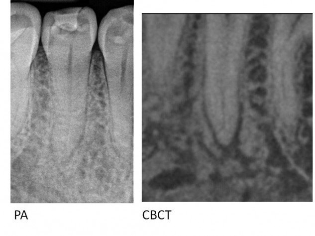
Premolars often have wild anatomical variations in their pulpal anatomy. Not only do we need to locate and adequately clean this anatomy for successful treatment, but we need to do so without needlessly weakening the tooth. Premolars will often branch from one to two or three canals. When they do this in a single-rooted tooth, there will often be deep concavities on the external root surface. These concavities mean that the dentinal walls surrounding the canals are thin in this area, and strip perforations are very common when “standardized” shaping techniques are used.
Pre-operative CBCT evaluation can be used to identify these risky cases, and root-form appropriate cleaning and shaping techniques can be used to conservatively locate the secondary anatomy and avoid iatrogenic errors.
I treated this tooth in November of 2011. Pt presented with class III mobility, with the chief complaint of being able to depress the tooth. This tooth was so loose that the patient felt that it would fall out. Initially, a periodontal probe could reach the apex of the tooth. At recall, we can see that osseous regeneration is complete (on the PA radiograph and CBCT images). Mobility and periodontal probings have both returned to normal.
Many teeth are needlessly taken out that are savable with endodontic treatment. Part of the problem is that we are conditioned to think that a certain look is an appropriate gauge for biologic success. A second reason is that dentists don’t feel comfortable treatment planning around complex cases, as the outcome is less predictable.
This patient presented with 9/10 pain from the apical periodontitis associated with the post perforation in the distal root. After the post was removed, canal negotiated and cleaned his symptoms completely resolved. In order to gain treatment planning predictability, the case was monitored (and re-cleaned) until bone-fill was evident. The 1.5-year recall shows complete healing (note regeneration of a continuous PDL space and lamina dura). Notice that the mesial roots were not negotiable on the re-treatment, but healing still occurred since the biology was treated, as opposed to the radiographic esthetics. This patient was very happy to predictably save his tooth.
On average, the minor foramen is 0.5 mm coronal to the major foramen, which is 0.5 mm from the radiographic apex (Kuttler 1955). Gutta percha cannot seal beyond the narrowest diameter of the root shape, or into a divergent apex(Weine 1975). Gutta percha beyond this point certainly won’t help, and although tolerated by tissues (Seltzer 1975), extruded filling material is associated with delay healing (Molven 2002). Cone Beam Computed Tomography can help to determine where the actual minor constriction is in relation to the radiographic apex. This way, treatment can be more precise than “filling” every case to the radiographic apex to achieve “the look”, and deleterious effects can be avoided. Note how in the case above, the pre-op PA does not indicate where the minor constriction is. The CBCT view shows that the minor constriction is 3.2 mm coronal to the radiographic apex. The case was filled to the minor constriction, and excellent healing is evident at the 1-year recall.
This case follows the premolar post below, and highlights the importance of treating complex endodontic anatomy. Leaving canal branches untreated can lead to endodontic failures. When retreating a case like this, it is beneficial to locate the missed anatomy. Shaping a tapered apical preparation will aid in the disinfection and obturation as well. Warm vertical compaction techniques help to obturate these spaces when the heat source can be carried down to 3mm from the canal terminus.
Below is another retreatment case, where the original RCT failed because apical anatomy was not addressed. It is an example of how CBCT helped to determine that an apical perforation had occurred, and the location of the actual canal. Again, failing to treat this complex apical anatomy contributed to the original treatment failure.
Every tooth is important. Some teeth may carry more weight than others. For example, the tooth in this case is the distal abutment for a three unit bridge, and the posterior left occlusion hinges on whether or not this tooth can be saved.
Treatment planning these case types can be challenging for the general dentist because the reported success rates for endodontic retreatment are lower than for initial treatment. Furthermore, this tooth has a 7mm probing depth and lateral radiolucency on the mesial surface of the mesial root. Combined with this tooth being a distal abutment, this tooth is highly suspicious of having a non-restorable vertical root fracture. The pre-operative prognosis is guarded.
The treatment planning options for this tooth include:
1. Endodontic retreatment, saving the existing bridge.
2. Extraction, without replacing the teeth, leaving no left molar occlusion.
3. Extraction, replacing occlusion with a removable partial denture.
4. Extraction, replacing with two implants, two implant crowns, and a new crown on # 20.
Obviously, the best option is to save the natural tooth with endodontic retreatment, assuming we can be assured of a favorable prognosis. This option is the least invasive, most functional, and a fraction of the cost of replacement with dental implants.
Treating teeth with a questionable prognosis in multiple visits allows us to evaluate healing prior to completing the case. Confirming that a case will heal prior to finishing takes the guesswork out of treatment planning around questionable teeth. For this case, we were able to confirm success by confirming that the perio probing depth healed to 3mm, and osseous regeneration has occured. Now, treatment planning is straight forward. The tooth has been saved, so we can retain the tooth and the existing bridge.
In my office, if we cannot save the tooth in question, we do not charge the patient for the treatment, thus eliminating the financial risk for the patient and referring doctor. There is no additional fee for this service.
This case illustrates how modern technology, modern techniques, and new materials can be used to save teeth that would have been slated for extraction in the past.
This patient presented with a previously root canal treated tooth with iatrogenic perforations and an untreated MB2 canal. It became symptomatic, with a chronic apical abscess.
Cone Beam Computed Tomography aided in locating two perforations in this tooth, that occurred during the original treatment. The first was a strip perforation in the MB1 canal into the furcation. The second perforation was in the palatal canal, and a gutta percha cone was extruded through the perforation. CBCT also helped to locate a previously untreated MB2 canal.
The surgical operating microscope allowed for visualization of the perforations and the MB2 canal during all phases of treatment.
Endovac negative pressure irrigation was used to clean and disinfect the perforations.
Mineral Trioxide Aggregate allowed for predictable repair of the perforations.
The patient was very happy to save her tooth from extraction!
Although the literature may show that there is no difference in treating cases in one vs. two visits, I almost always treat cases with symptomatic apical periodontitis in multiple visits. The main reason is for predictability: I can make sure that we are able to resolve the symptoms prior to finishing the case. A second reason is that we can use calcium hydroxide to help disinfect the root canal system. In the last few years, technological advancements in irrigation have been shown to increase the efficacy of irrigants at disinfecting canals. For patients who have difficulty tolerating dental treatment, the potential exists to treat these case types in a single visit.
Above is a two year recall of #12. The tooth initially presented with a necrotic pulp and an acute apical abscess (acute exacerbation of chronic apical periodontitis, or “Phoenix Abscess”), and a buccal space infection. EndoVac apical negative pressure irrigation was used and treatment was completed in one visit for this 82 year old. Note that the canal shapes are still conservative (30-06). A fiber post was bonded into each canal. The two year recall shows success, with resolution of the periapical radiolucency, and the return of the radiographic PDL space and lamina dura.
In this case, an indirect pulp cap was done by the patient’s dentist years ago, and recently became symptomatic. Clinical photo of the complex microvasculature in a vital coronal dental pulp. This # 30 had five canals, with the fifth being a mid-distal. Endovac apical negative pressure irrigation is useful even in this case type. Although the main canals were only shaped to a 20-07 and the mid-distal to a 20-04, the microcannula reached beyond where the distal canals merged and allowed for a high flow of irrigants through each canal, to effectively remove vital tissue from the anastamosis. Fiber post bonded in the DL canal.
Tooth #20 presented with previous RCT and persisting Symptomatic Apical Periodontitis. The periapical radiolucency is bi-lobed, suggesting two portals of exit. It is important to locate and clean complex anatomy in order to heal the disease. The location of the “lobes” of the lesion give the location of the anatomy away, as the POEs tend to be centered in each lesion. I almost always complete re-treatment cases in multiple visits, to utilize calcium hydroxide as an inter-appointment medicament. There have been a few recent studies suggesting a good efficacy of negative pressure irrigation (the Endovac) in aiding canal cleanliness, even in one visit. This was my fist case using the Endovac in a one-step re-treatment. The shape was finished at 40-08, which is bigger than I would shape it to today. After debriding the accessory canal and flowing high volumes of irritant to the apex, the two year recall shows an excellent result with complete regeneration of the periapical tissue.
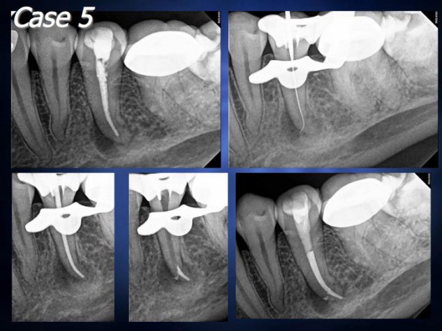
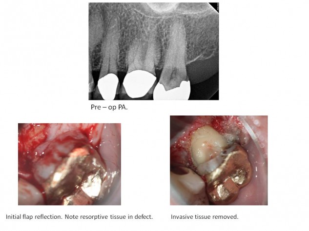
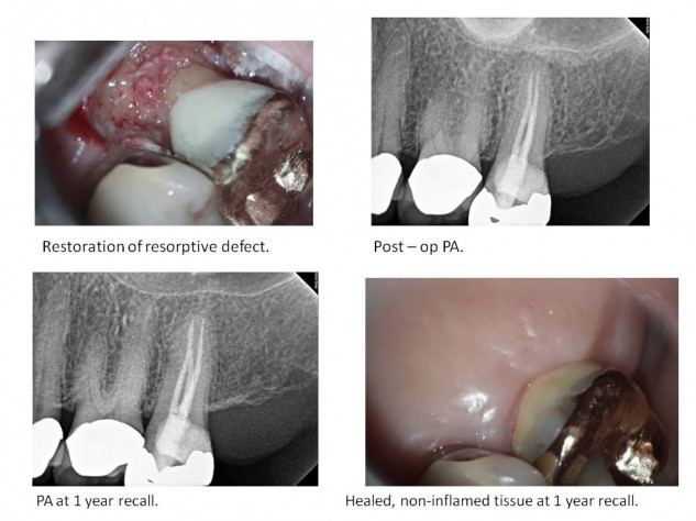
Patients are always happy to save their natural teeth. Even many teeth with significant defects resulting from External Cervical Invasive Resorption (ECIR) can be predictably saved.
ECIR is a non-inflammatory resorptive process that can occur when clastic cells have access to dentin following damage to the cementum / PDL protective layers of the root. After clastic cells remove dentin, PDL-like tissues invade the tooth. These lesions may become large because incipient lesions are difficult to detect, owing to the fact that they initiate within the periodontium, so are sub-gingival. The process is not related to the pulp of the tooth. Many cases are detected once the resorption reaches the pulp however, when the pulp becomes subsequently infected with oral microorganisms through the resorptive defect. At that point, patients will experience the typical symptoms of irreversible pulpitis.
The case below is an example of an extensive ECIR defect on the palatal of # 15. The patient’s chief complaint was lingering sensitivity to cold. A surgical approach from the palatal was used to debride and restore the area of resorption. Root Canal Therapy was completed because the pulp had become secondarily infected. When the patient returned for her one year recall, she reported that her tooth and gums were completely asymptomatic and functional, and she expressed her appreciation to be able to save her natural tooth.
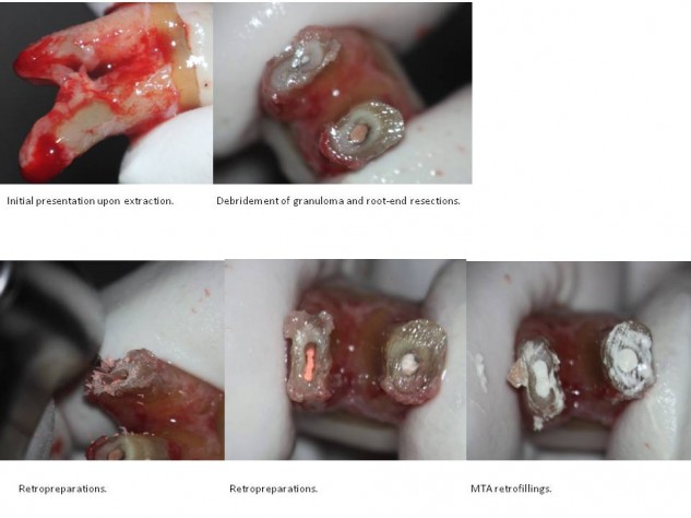
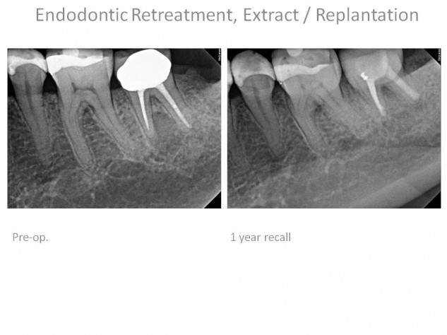
Much of the success of endodontic therapy is determined by the ability to remove pathogenic microorganisms from the root canal system. In most cases this can be accomplished through orthograde root canal therapy. When apical complexities that aren’t treatable from an occlusal approach harbor persisting microorganisms, they are often predictably treatable with a surgical approach. Second molars are often difficult to access surgically, which makes intentional extraction followed by root-end resection, retro-preparation, retrofill, and replantation a viable option.
The case below illustrates this case type. Endodontic retreatment was performed due to Symptomatic Apical Periodontitis. Patency was not achievable due to ledging / transportation from the initial treatment. Consequently, sysmptoms persisted following retreatment due to bacteria remaining in the apical anatomy. Rather than extracting the tooth and being left with an edentulous area or prosthetic implant, the root canal treatment was completed ex-vivo and the tooth was replanted back into it’s natural site.
One year recall shows complete osseous regeneration of the Apical Periodontitis lesion. This tooth is free of symptoms, without mobility and is completely functional. The patient was extremely happy to be able to keep her natural tooth.
Perforation of a tooth during access used to have a poor prognosis, and often led to the extraction of the affected tooth. Fortunately, with modern materials and techniques teeth with perforations can now be predictably saved.
This case was started by a dentist, using the classic access design from the palatal toward the facial. An iatrogenic perforation was created through the facial aspect of the cervical third of the root while searching for the calcified canal. The sagittal view of the cone beam image demonstrates how this orientation for access will inherently lead to this type of perforation. The sagittal view also shows how the more appropriate straight line access is right through the incisal edge.
This tooth was treated in two visits. The perforation was repaired with white MTA, a glass ionomer orifice barrier was placed, and sodium perborate was sealed in the access as a walking bleach technique. An access through the incisal edge in the direction of the long access of the root was used to locate and treat the calcified canal on the second visit.
When a coronally loose #10 K-file won’t slide the last couple of millimeters to patency, as seen in the WL radiograph, it is almost never due to an apically calcified canal. If a hard, abrupt impediment is felt, it will either be the outer wall of a curve or an apical split. DB roots of maxillary molars are notorious for apical dilacerations to the distal. In this case, the file is hitting against the outer wall of the apical curve in the WL radiograph. Patency was achieved with a pre-bent #6 K-file (UR rad, squint), and the shape was completed with hand files, including a pre-bent GT 20-06 rotary hand file. A pre-bent master cone was fit to length and obturation was completed.
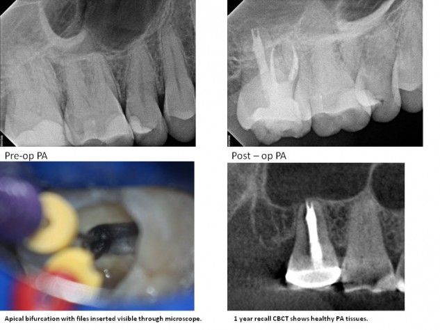
This patient presented with Symptomatic Chronic Apical Periodontitis resulting from a necrotic tooth # 19. Our ability to resolve Apical Periodontitis is dependent on our ability to disinfect and seal all aspects of the root canal system. This tooth presented with severe calcification of both the mesio-buccal and mesio-lingual canals, which makes locating and treating these canals particularly challenging.
Root canal systems in mesial roots of lower molars are never straight. From the pulp chamber, going apically, the canals begin in the mesial direction and curve around to exit towards the distal in most cases. This curvature makes deep troughing for a patent lumen in a calcified canal risky. Since “drilling” is only possible in a straight line, any specific morphologic information obtainable is critical to decrease the chances of iatrogenic perforation or excessive gouging while attempting to locate canals by troughing deep into the root.
Further complicating this case type is the lack of anatomical landmarks. Natural extra-coronal landmarks are lost when prosthetic crowns are fabricated. The intra-pulpal dentin map becomes less useful as troughing continues further down a curved root.
In this case, both pre-operative and inter-operative CBCT scans were indispensable in locating the highly calcified mesial canals. Since the distal canal was easily located, it was used as a landmark to find the others. The distance and direction to look for the ML in relation to the D was determined using the CBCT axial view at the first appointment. After locating the ML, and troughing 4mm into the MB to no avail, CaOH2 was placed and an interoperative CBCT was taken. This scan was then used to find the MB canal in relation to the previously located canals and the area already troughed. The crown was removed and a provisional was fabricated during the first appointment.
This approach allowed for complete treatment of the root canal system. Importantly, the CBCT guided troughing allowed for a conservative removal of tooth structure to locate the calcified canals, which significantly increases the long term prognosis compared to blind drilling and gouging.
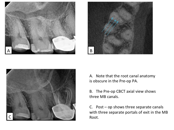
Although many text books will describe premolars as having one canal, anybody who practices endodontics will tell you that this is almost never the case. In fact, premolars can often be more difficult to treat than molars due to their anatomic variability and often skinny root and crown forms.
Maxillary second premolar root canal casts from Hess’ classic 1921 study:
Below are cases completed by Dr. Boehne demonstrating considerable variability in premolar anatomy. Note that the portals of exit in these cases range from one to eight. The number of canals are often variable within the same root as canals split and merge and re-split; note the 1:2:1:2 system in the upper right case. Root lengths and curvatures may differ vastly in the same tooth. Premolars commonly have apical bifurcations and even trifurcations and deltas. Contemporary endodontic techniques, and a lot of care, allow for the successful treatment of this challenging anatomy without the need for subsequent apical surgery.
The case below demonstrates a Root Canal Treatment that failed because the apical anatomy was not treated by the original treating dentist. Good osseous healing is evident at 10 months, after the apical bifurcation was addressed.
The Surgical Operating Microscope not only helps with locating and treating canals (primary endodontic anatomy), but it can also be a useful tool for locating and treating secondary endodontic anatomy. The case below demonstrates a deep apical bifurcation in the palatal root of # 2. This root was relatively straight, allowing for direct visualization of the bifurcation with the magnification and co-axial lighting provided by the SOM.
This 11yof had a traumatic fall, resulting in a complicated crown fracture on tooth # 9 and an uncomplicated fracture on # 24. One day of pulp exposure following the fracture caused Irreversible Pulpitis in # 9, and # 24 tested WNL to pulp sensibility testing. RCT was completed on # 9, and the tooth was restored with a fiber post and an esthetic bonded composite restoration. Tooth # 24 was restored with composite, and the occlusion was adjusted to light centric contacts without excursive interferences.
A “fast break” is a term used to describe a situation where a root canal disappears on a radiograph as you move apically. This happens when the main canal splits into multiple smaller canals that are not discernible on a radiograph. CBCT axial views are indispensable determining the number and location of these canals. Tooth # 13 (seen at the top of the CBCT axial view) branches into three canals. Note how a conservative access can be used to locate even multiple canals when their location is known prior to starting the case, significantly increasing the long term prognosis by preserving peri-cervical tooth structure.
The axial view of the CBCT image is extremely beneficial for determining the number of canals in a tooth. Even minor anatomy can be visualized on some cases. In this particular case, my assistant was able to identify the MB3 canal as the image loaded. Treatment of this tooth with a supernumerary canal was straight forward and predictable following pre-operative case assessment utilizing CBCT.
A “fast break” is a term used when a root canal seems to disappear on a radiograph as you move apically. It is often mistaken for a “calcification”, but in reality these are areas where the main canal bifurcates into multiple smaller canals that are too small to be discernable on a radiograph. CBCT can be useful to verify the location of this secondary anatomy. Note how on the PA, the canal is not discernable in the apical 2mm, whereas on the CBCT both apical canals are evident. CBCT technology facilitated the location, shaping, cleaning and filling of this complex anatomy.
A 31mm second molar with anatomy that makes both shaping and filling the root canal system a challenge.
Testimonials
Dan, Thank you so much for taking such great care of me! I’m super grateful for your generosity and my tooth feels great!
Todd M. San Clemente
Dear Dr. Boehne,
I want to thank you for the skilled and compassionate services you rendered to me with regard to my root canal. I was greatly impressed by your professional skills and your sensitivity to your patients’ concerns and fears. The kindness of all of your staff and the state-of-the-art equipment in your office, are also eloquent manifestations of your commitment to the best possible care of your patients. I am profoundly grateful that Dr. Franke sent me to you.
Sincerely Dwight A. Dana Point
Dear Dr. Boehne and Chelsie, I can not begin to put into words how much I appreciate what you’ve done for me. God is good. I’m glad my tooth doesn’t have a hole in it anymore. haha I wanted to paint you guys this so you would know how much what you did meant to me; and also so you guys could say you Do have an ocean view from your operatory. You guys are great!
Thanks again
Dear Dr. Boehne,
Thank you for treating a panicked old lady with kid gloves. It was greatly appreciated.
sincerely,
Judy, San Clemente
Dr. Boehne & staff,
I just wanted to thank you so much for your time and effort to fix my tooth! I really appreciate how you took the time to explain everything to me and make sure that it healed. Everything looks and feels great!
Thanks again,
Lindsay C, Newport Beach
Thank you Dr. Boehne and the team. I was treated with professionalism and I was well informed during the entire process. A truly fantastic experience!
Chris L.Newport Beach
Dear Dr. Dan and Ruthie;
I thank you, not only for your professional expertise and abilities, but also for the caring concern that you extended towards me. It turned what could have been a trying anxious time into a time of pleasant healing and nurturing. Thank you both.
With gratitude Sally F. Laguna Beach
Dear Dr. Boehne,
Waking up to sudden severe nerve pain and being told you need a root canal is not typically the way one plans to begin their day! However, from the moment I stepped into your office I felt I was in good hands. Your receptionist and your dental assistant were kind and reassuring while I waited to be seen.
I am a nervous dental patient and was unsure about the details of the procedure, but you took the time to explain very clearly and specifically what I could expect. You we… Read more
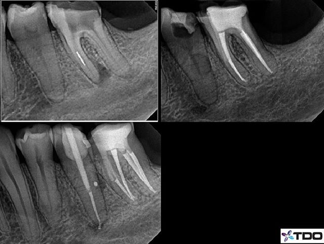
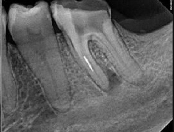

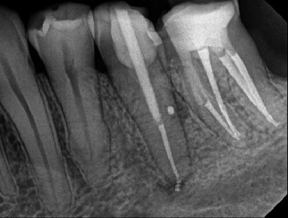
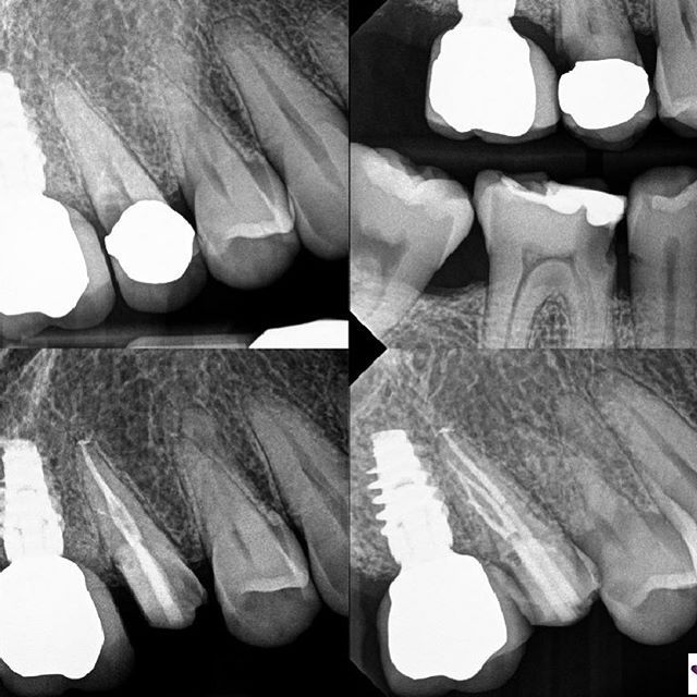
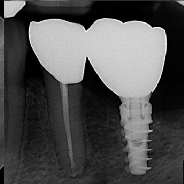
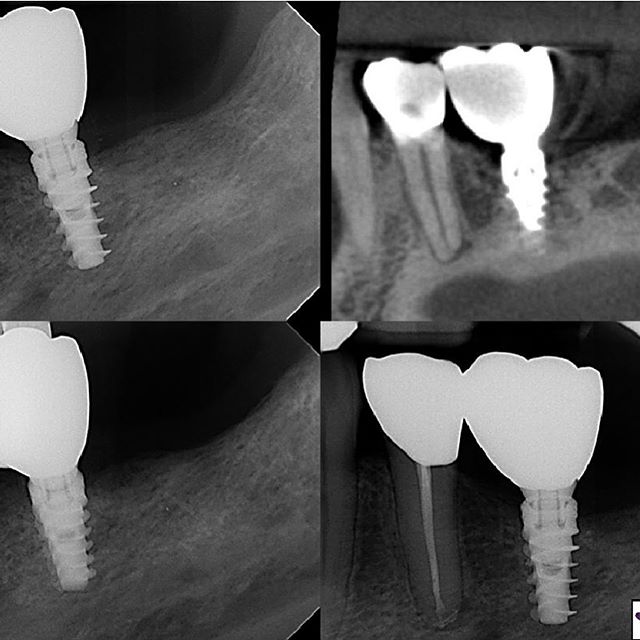
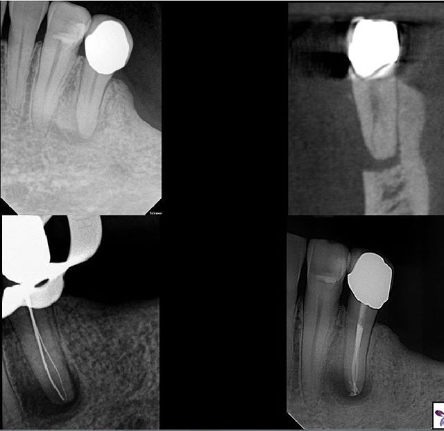
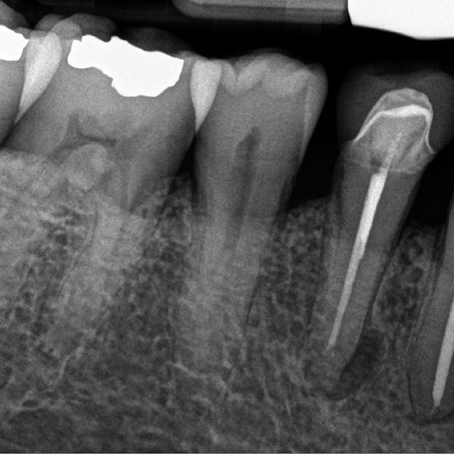
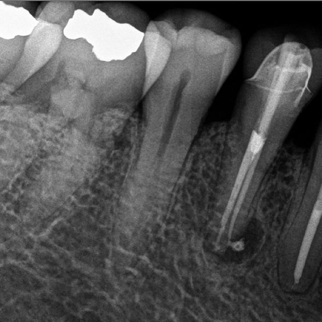
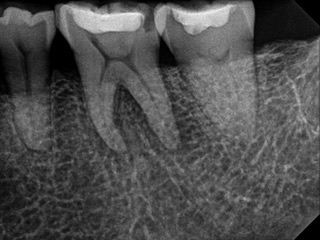

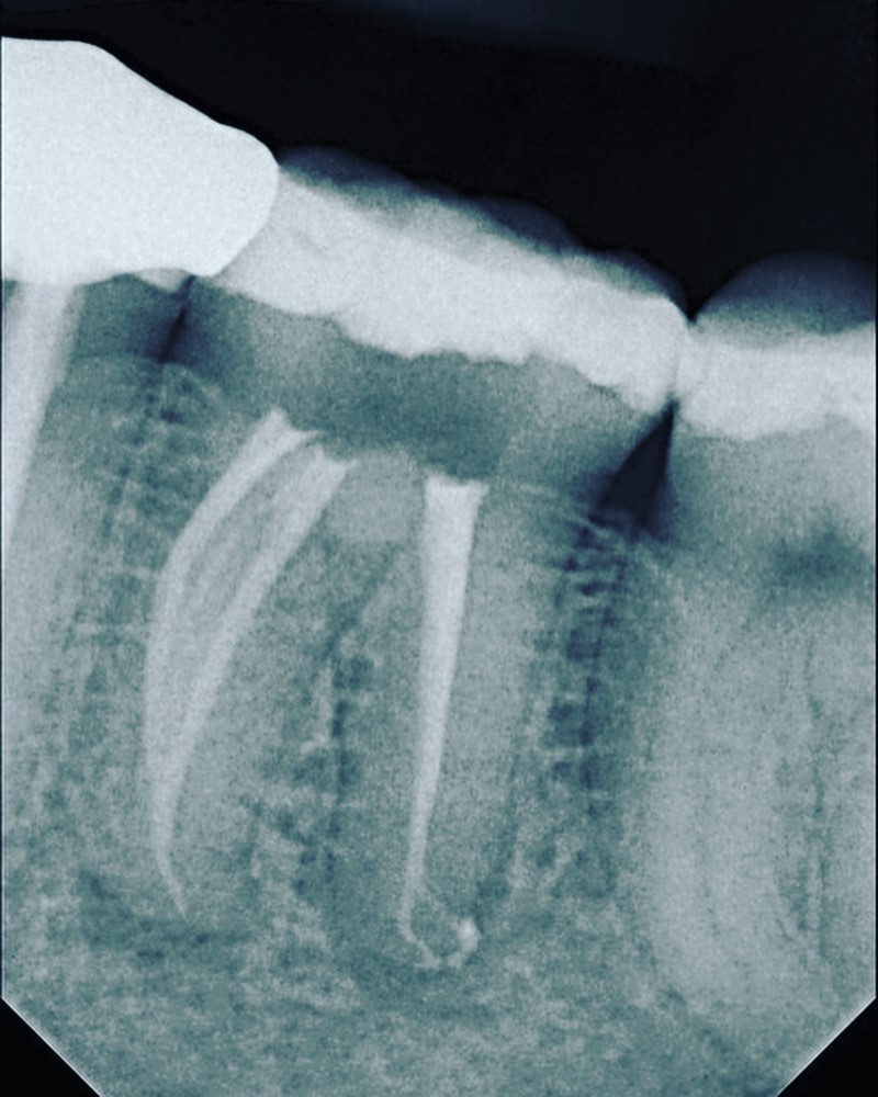


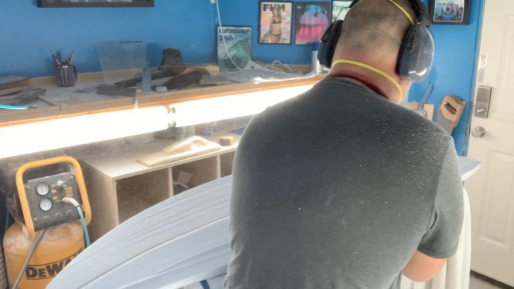

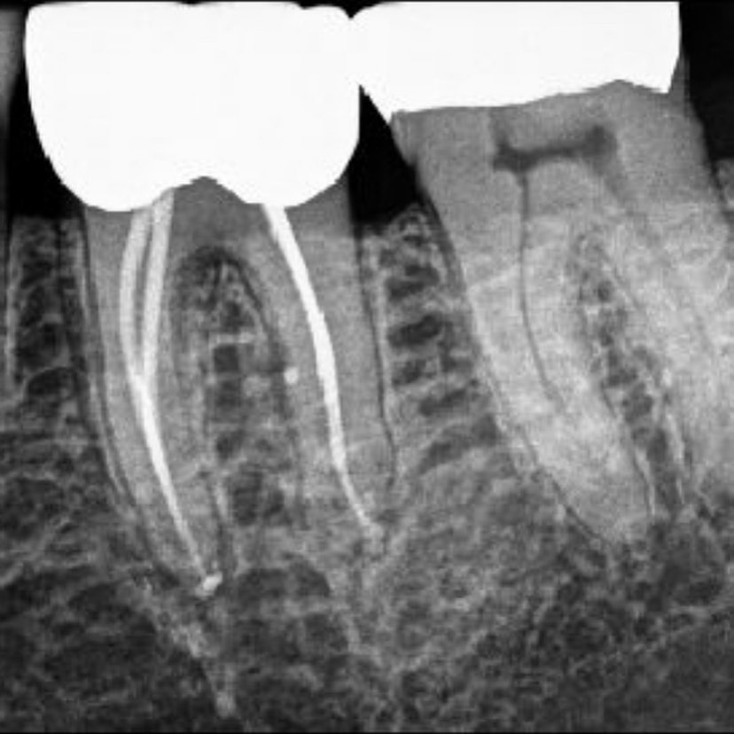

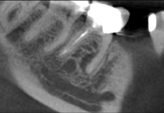


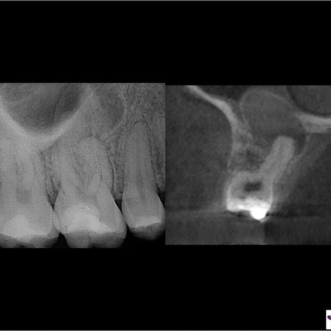


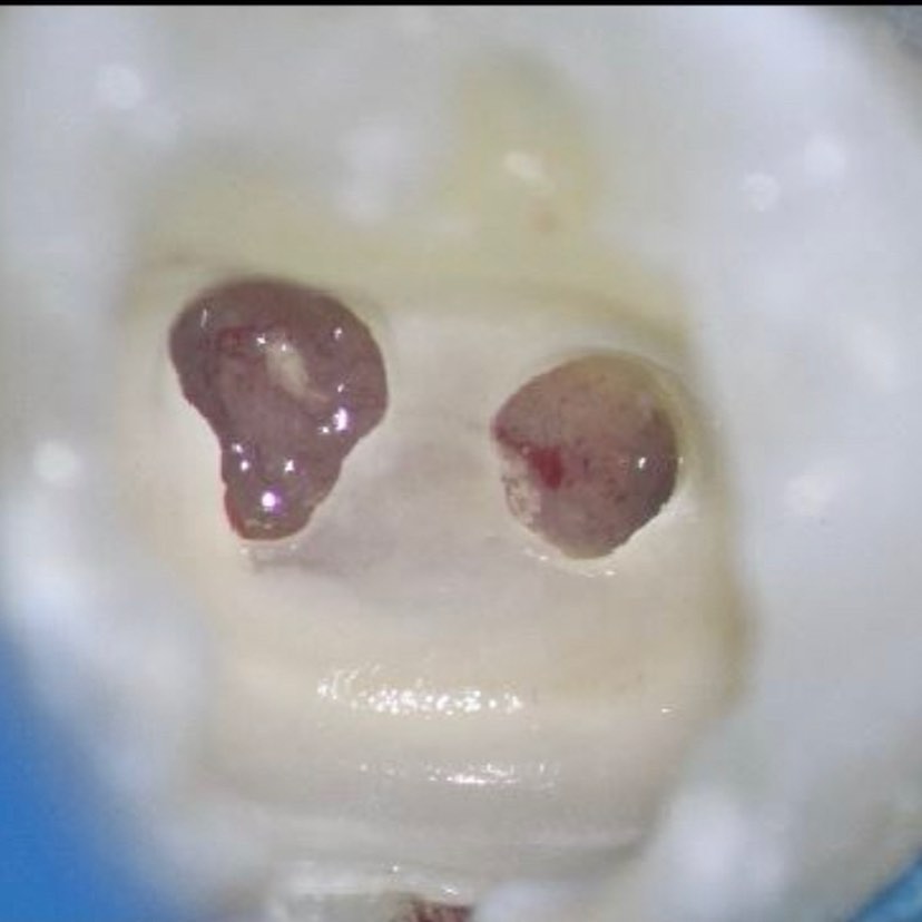


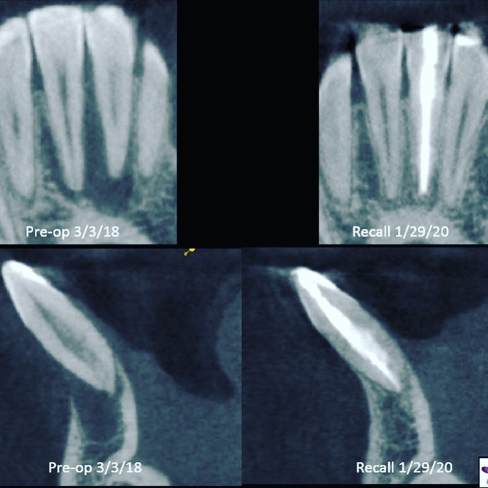


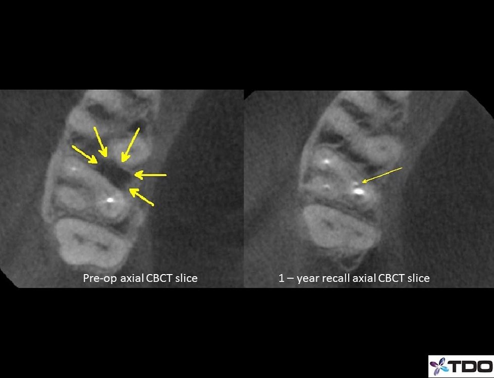
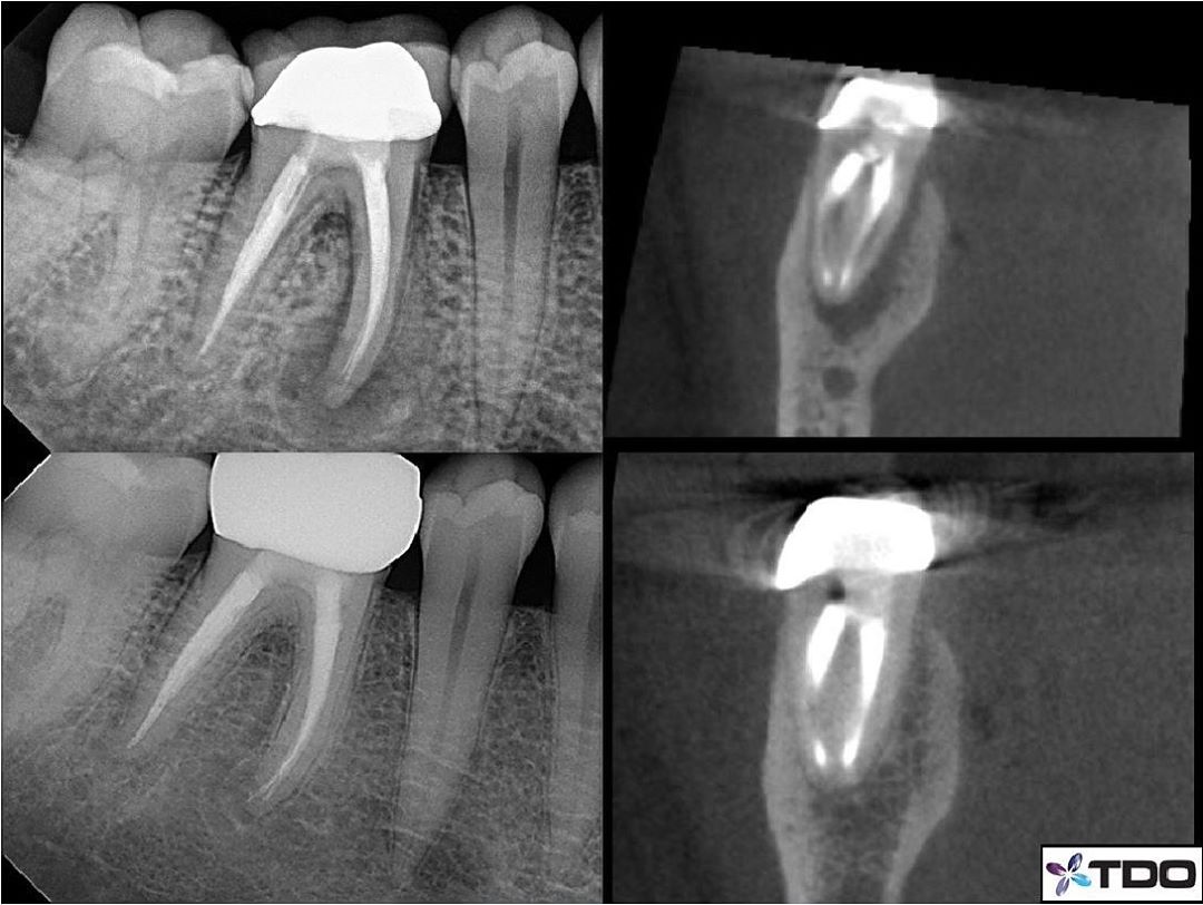

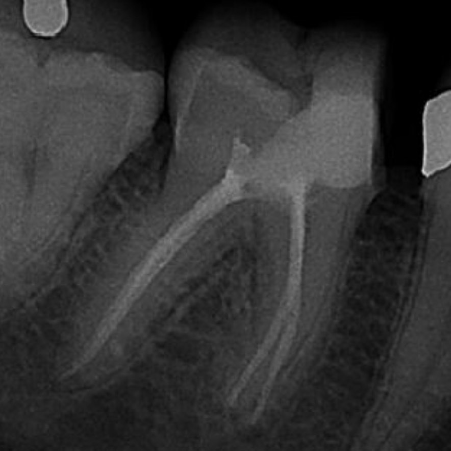

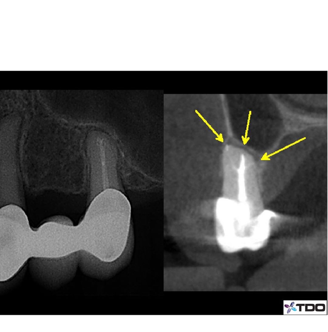


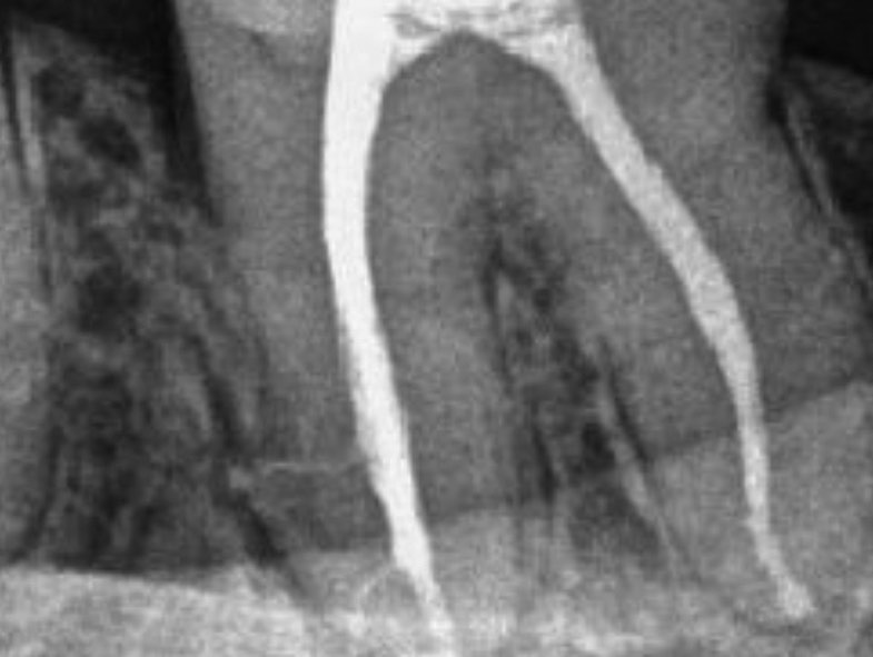


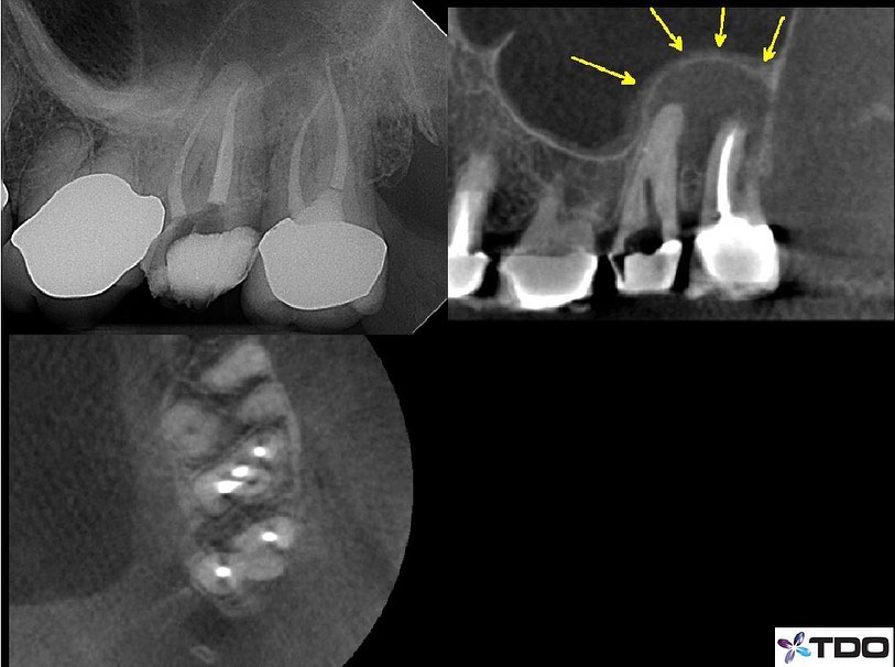


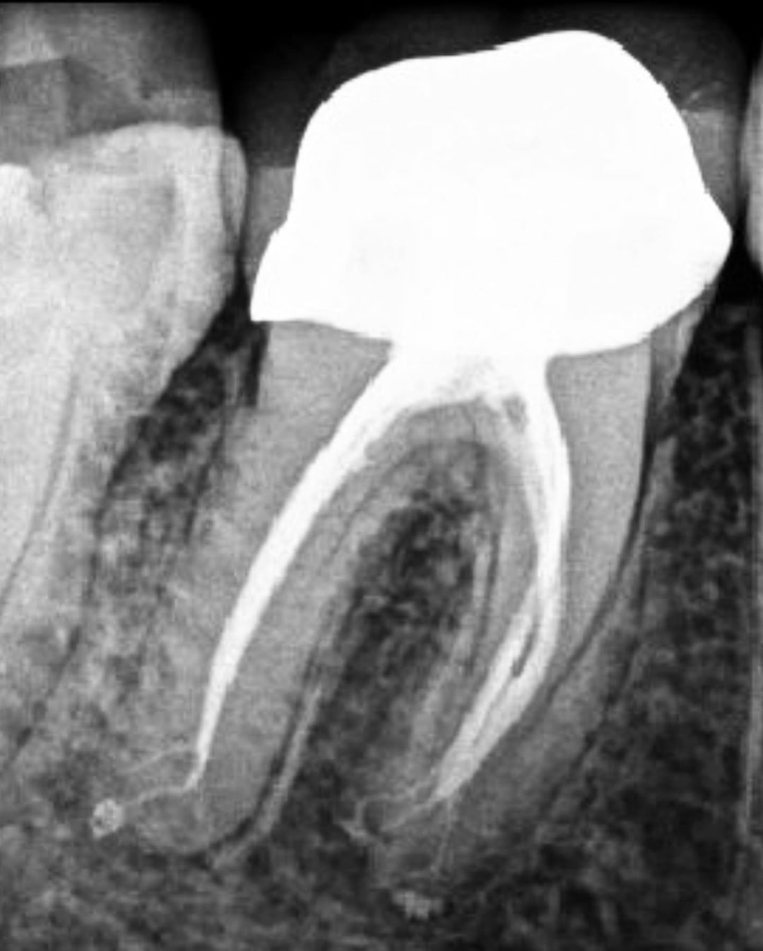


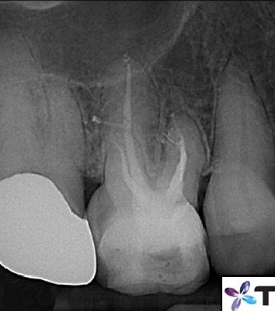

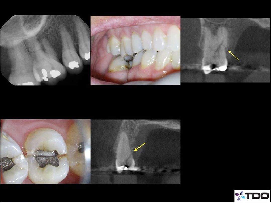


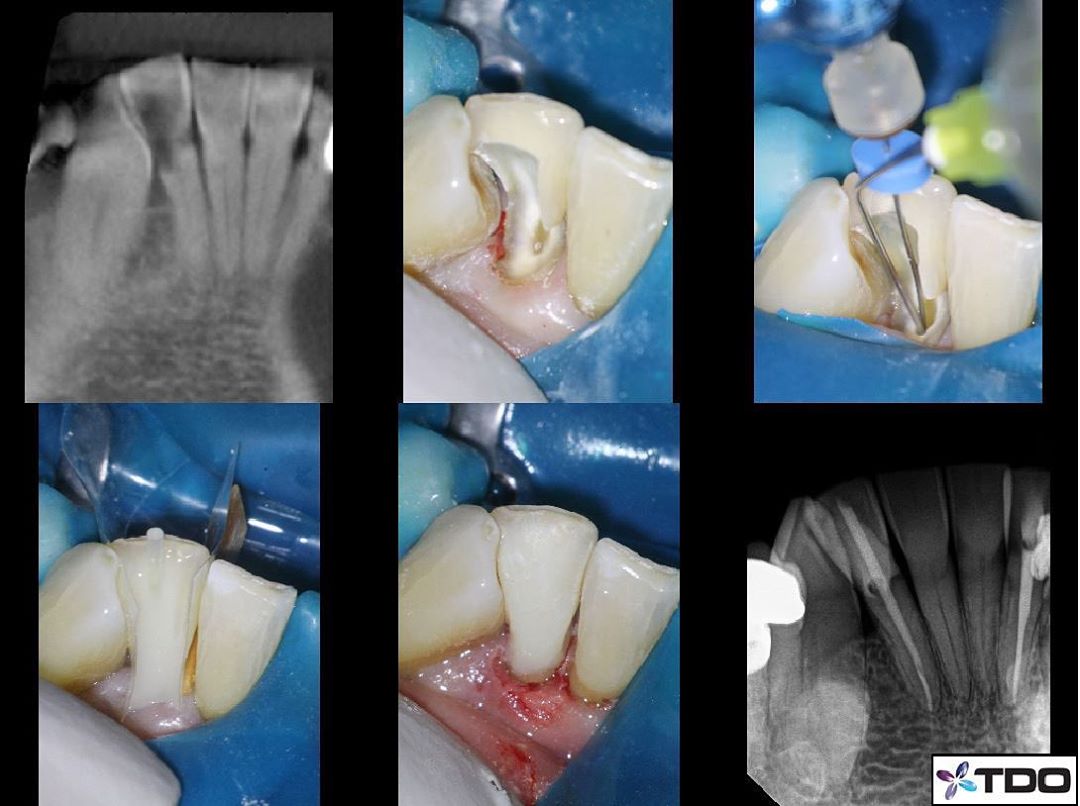

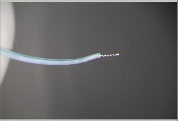


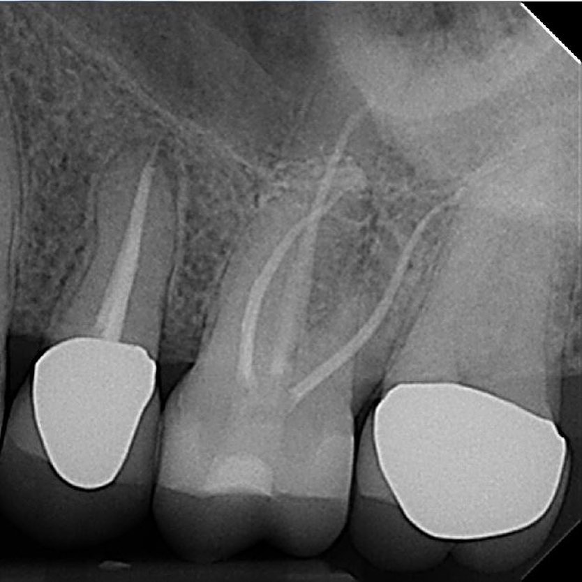


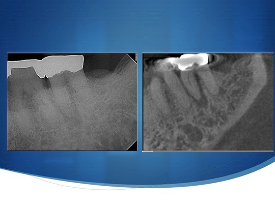

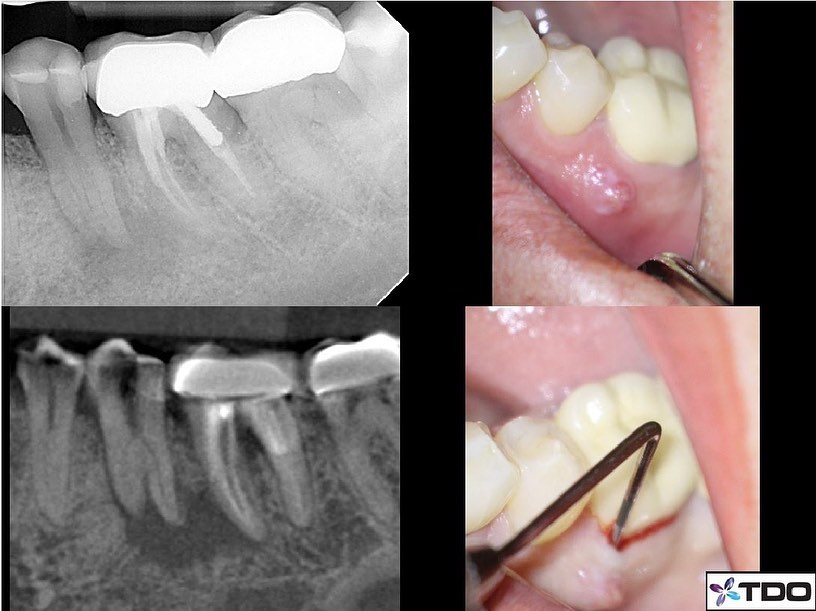

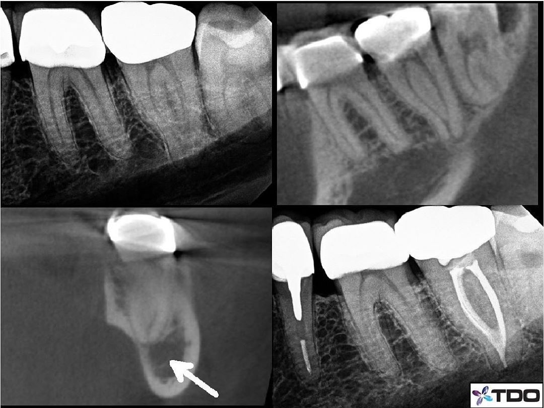


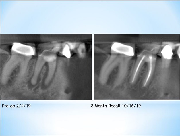
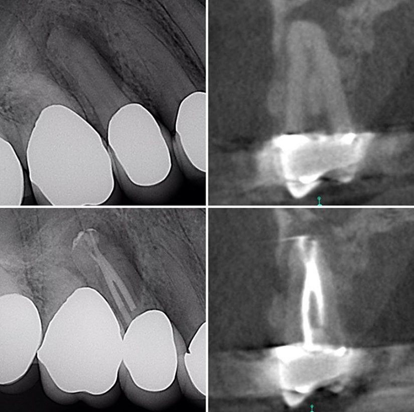


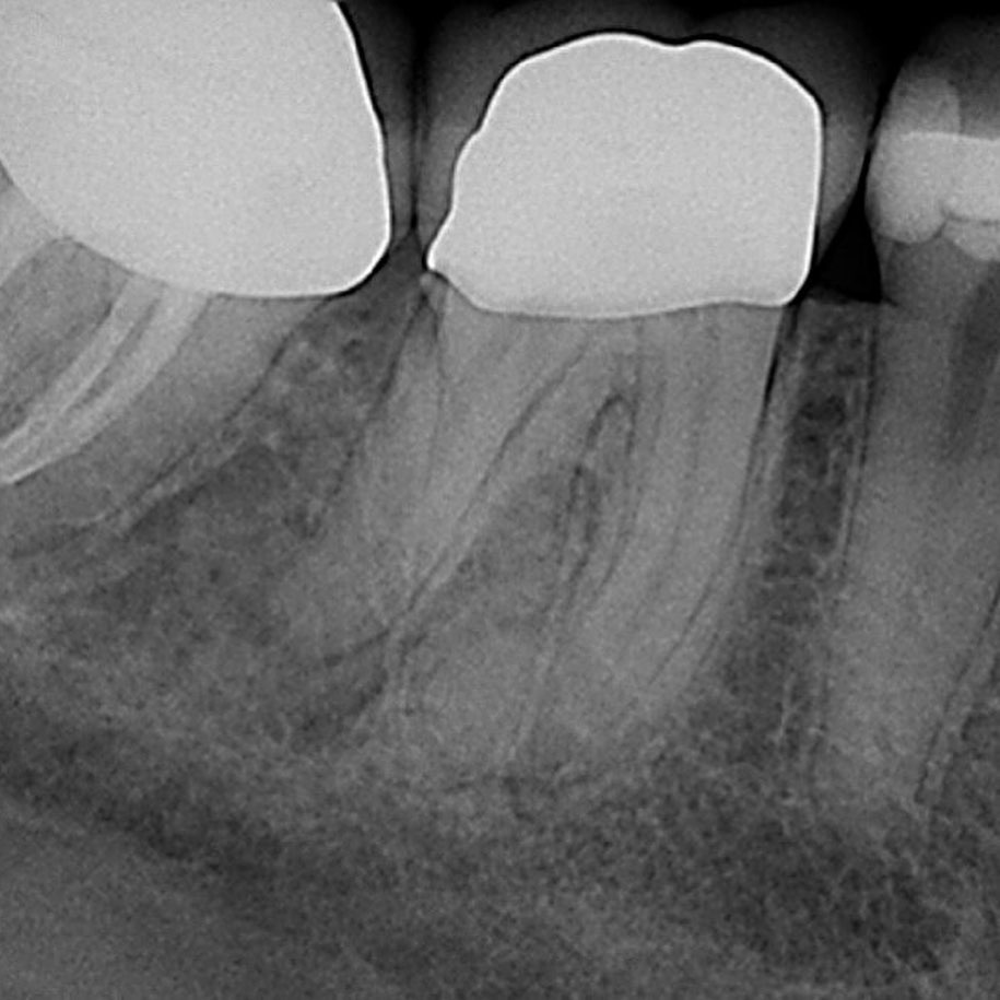


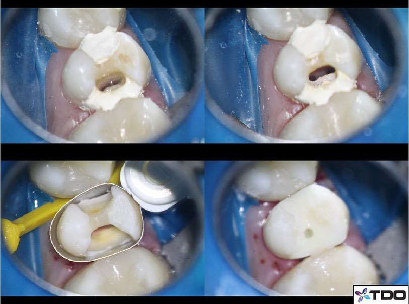

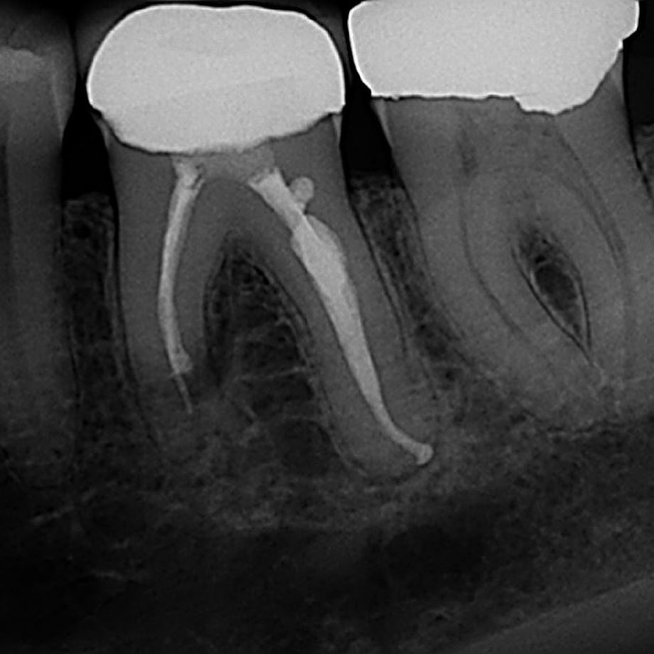


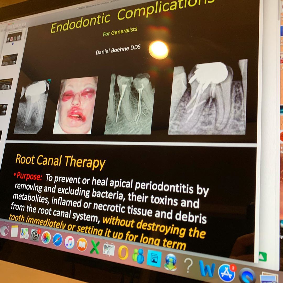


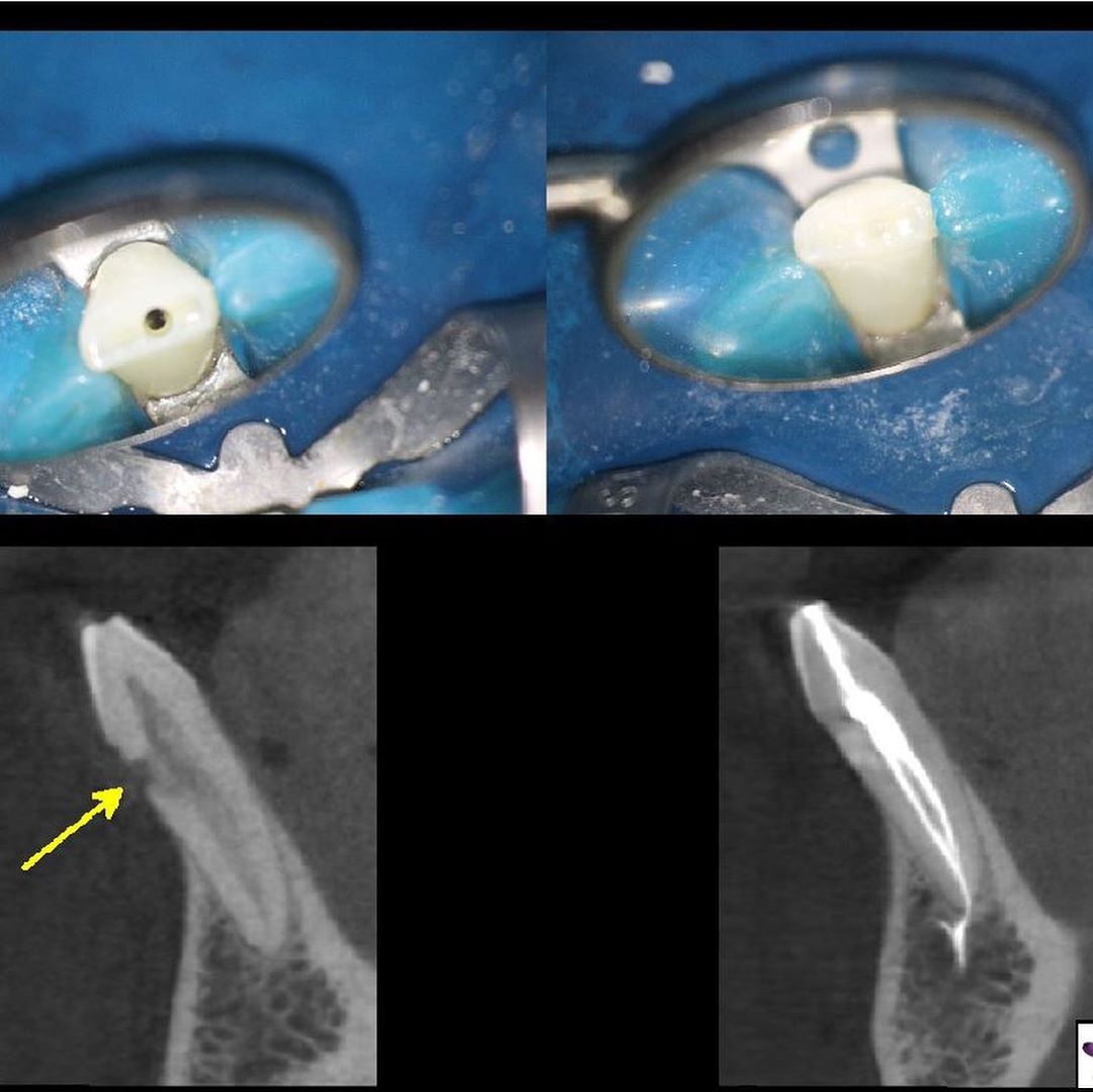
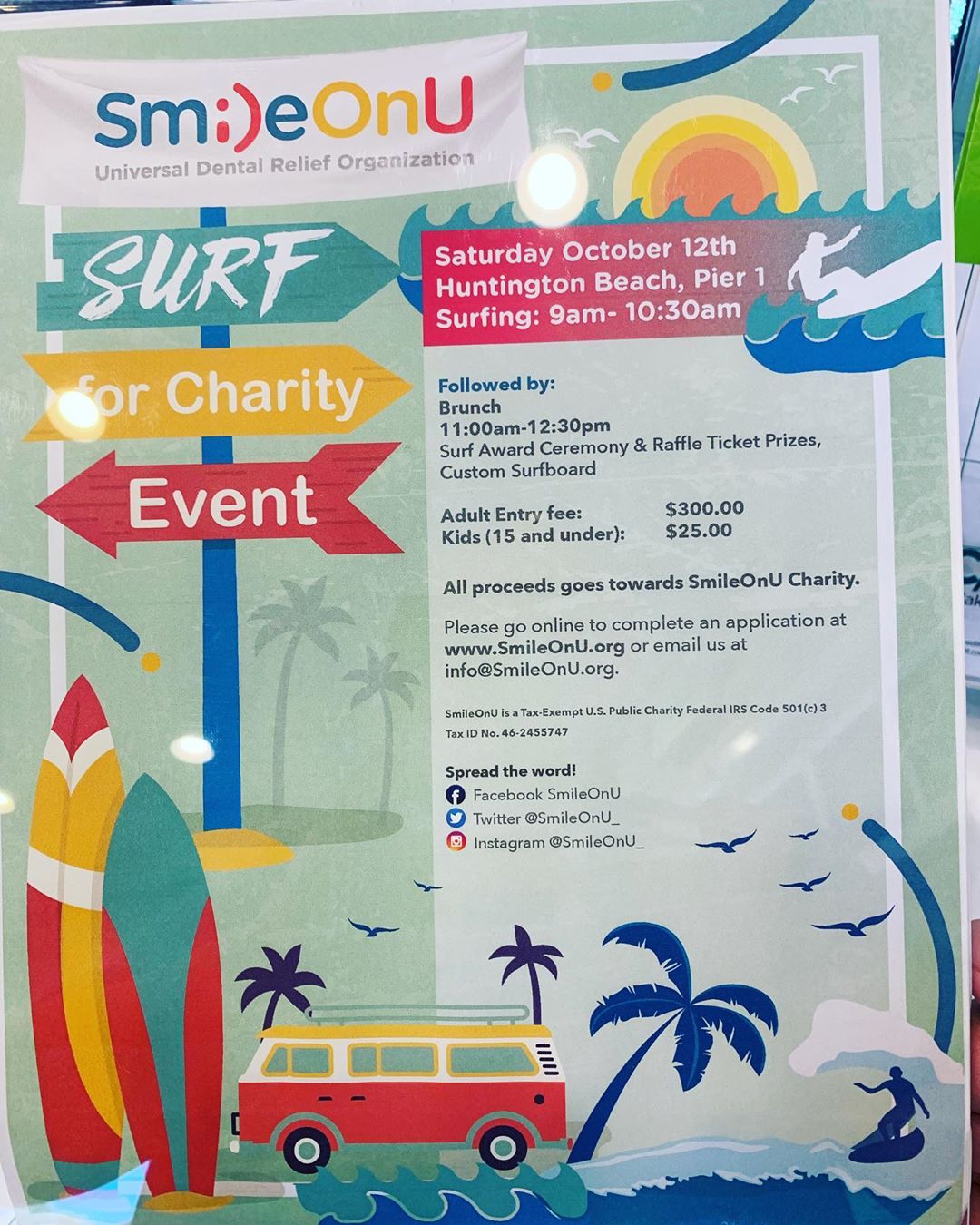

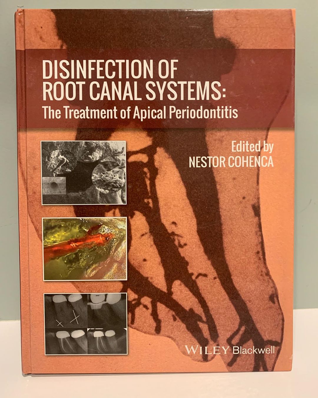


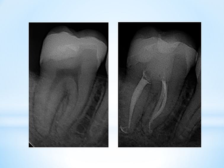
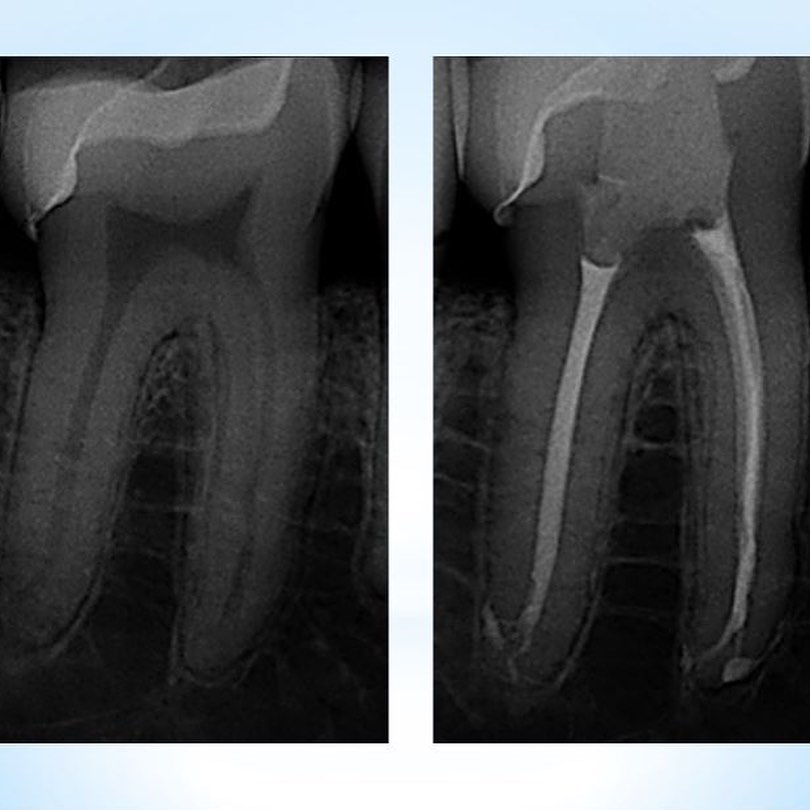


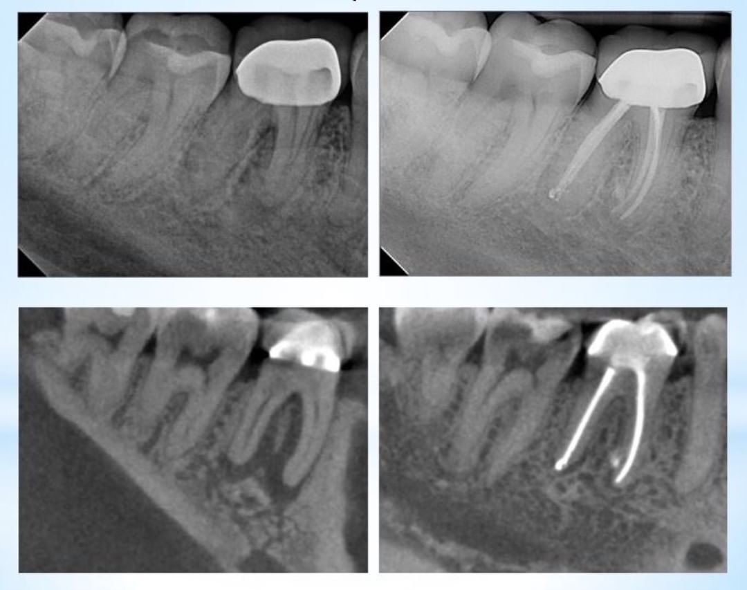
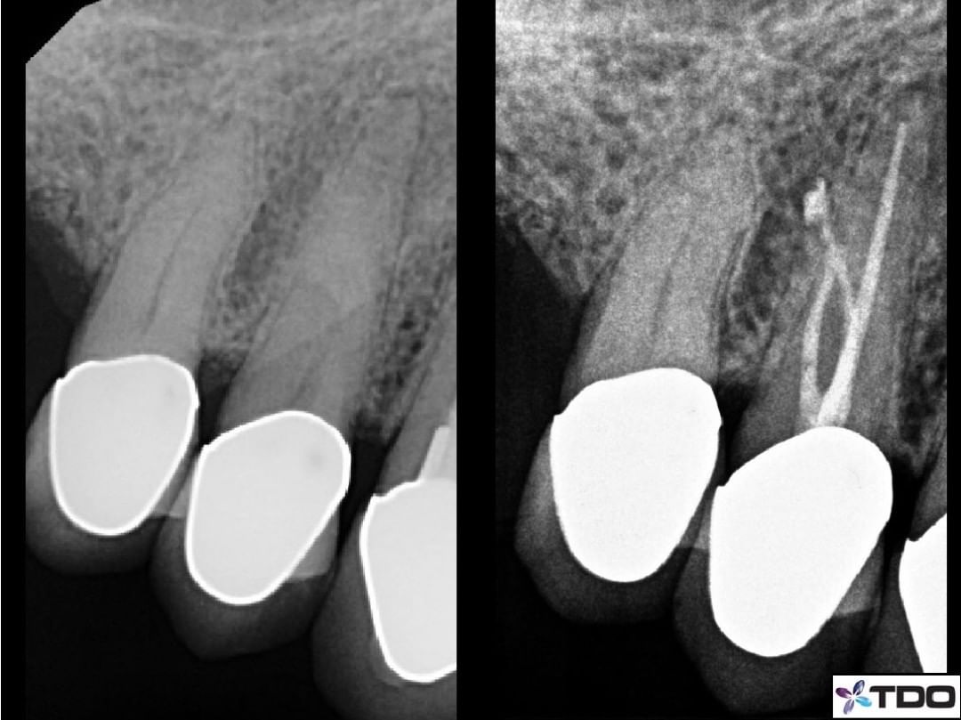
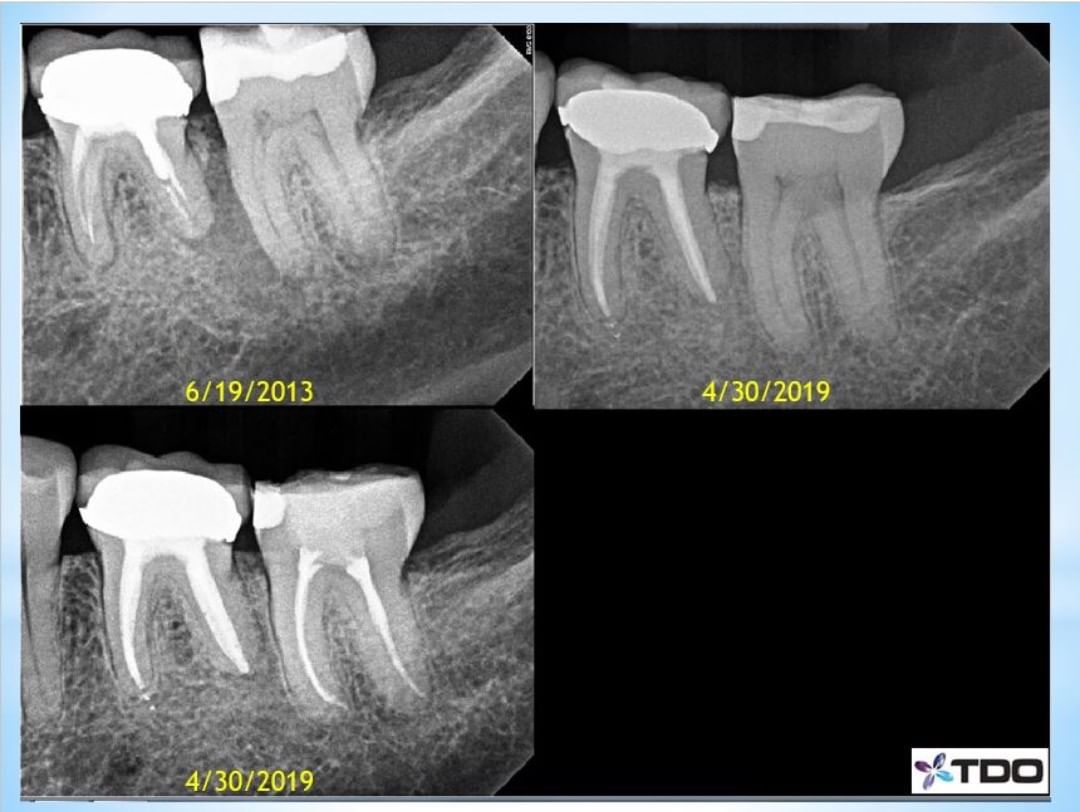
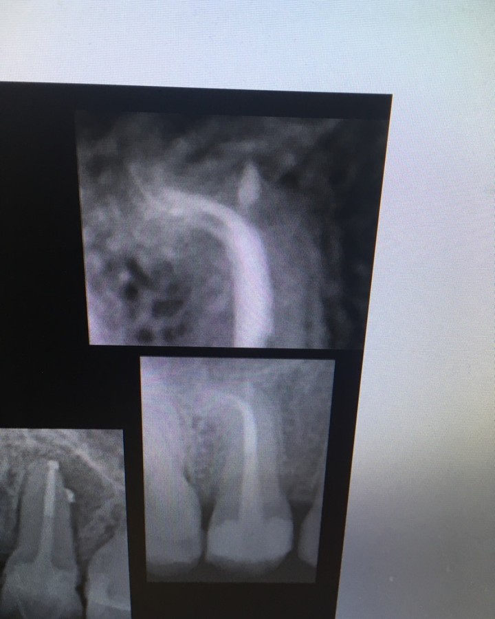
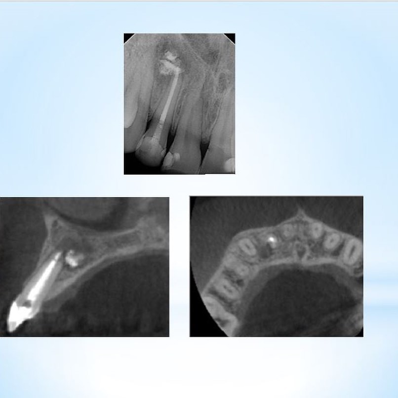


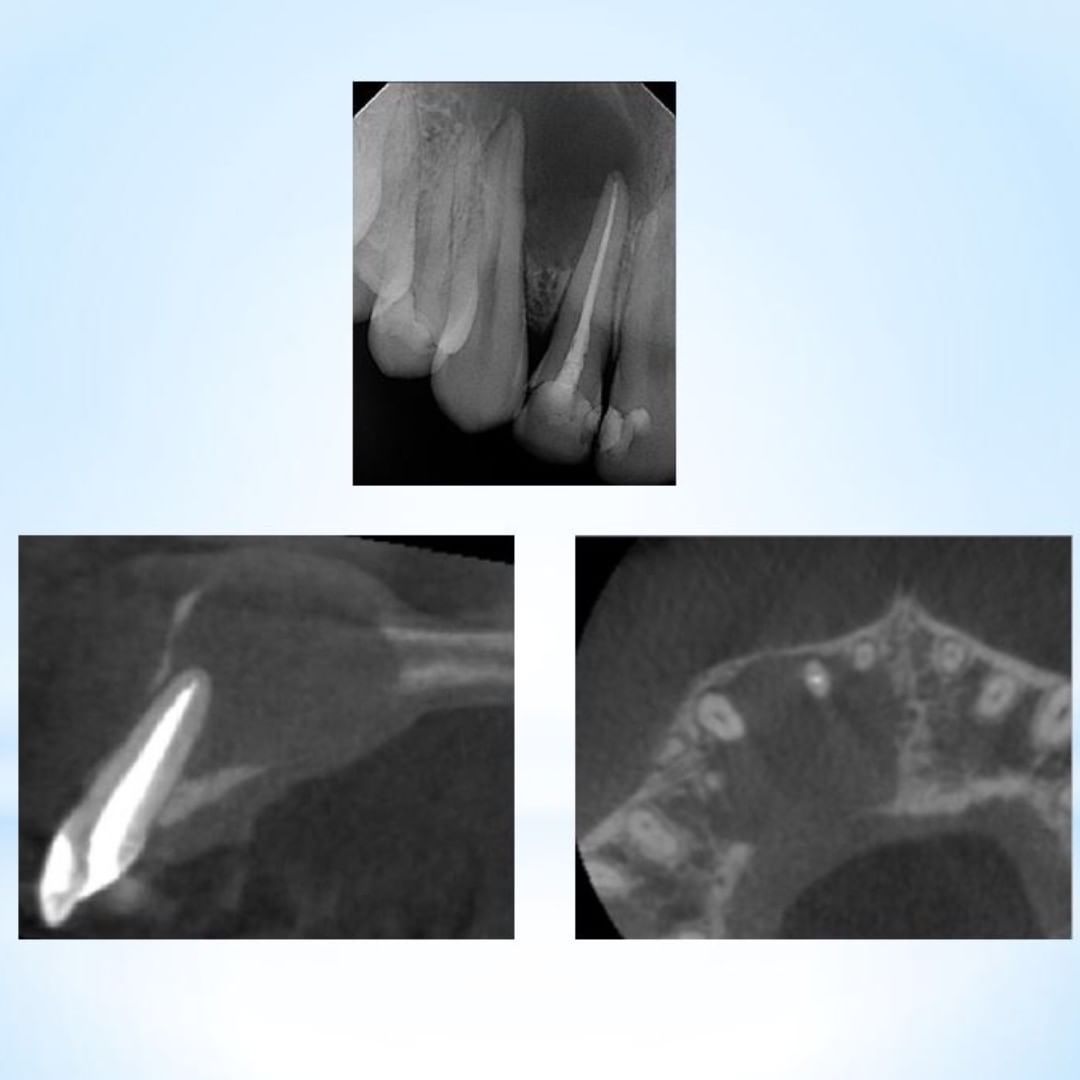
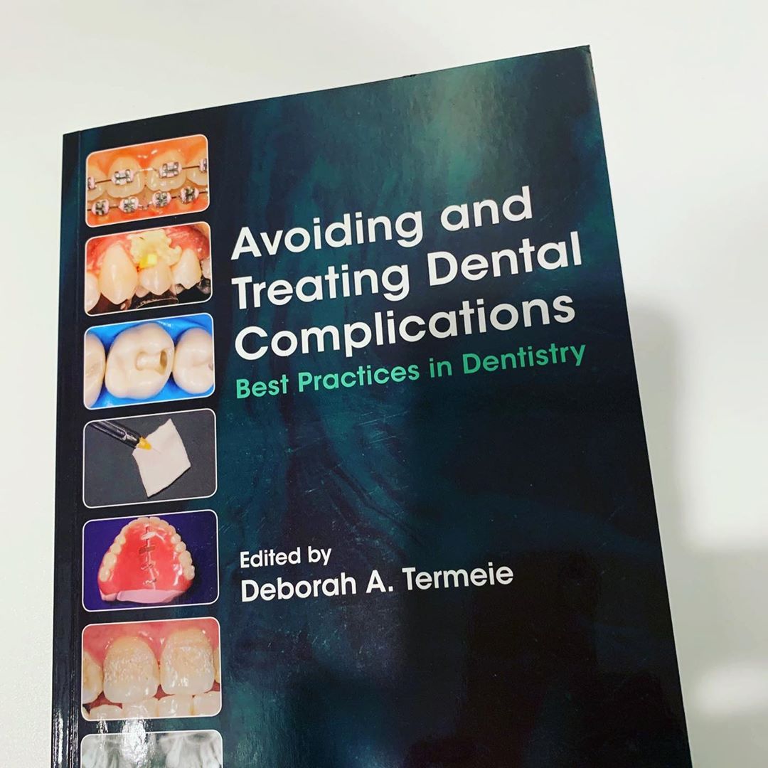

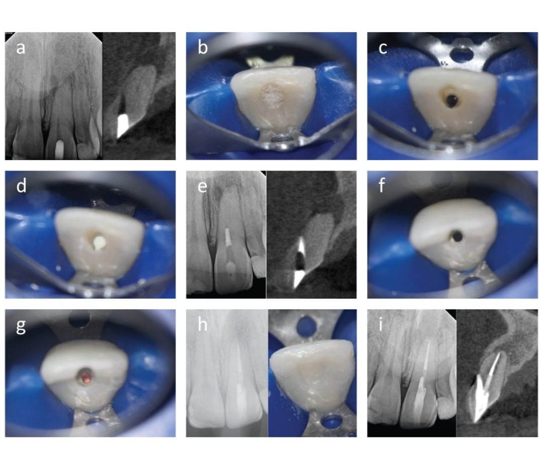
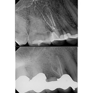

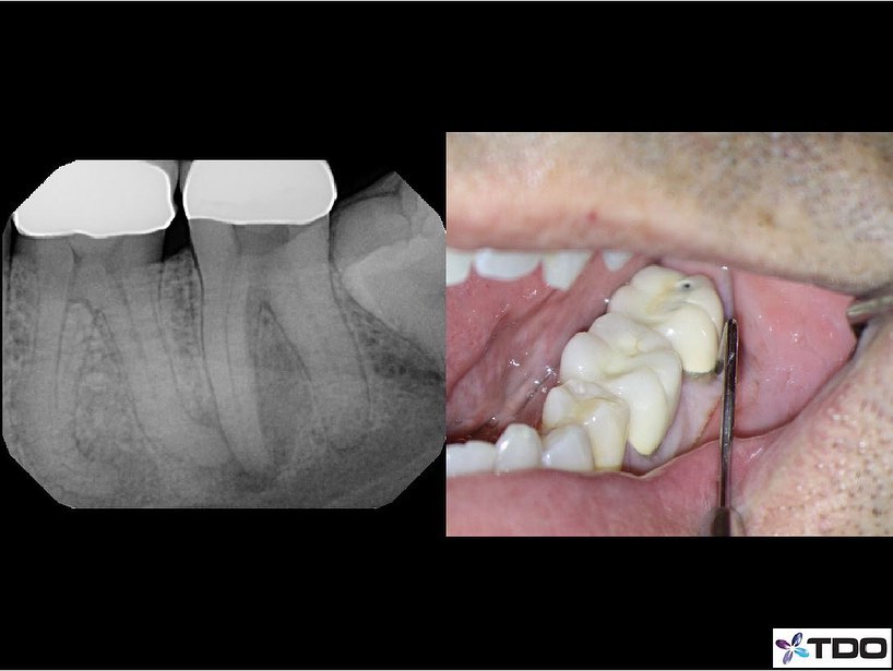


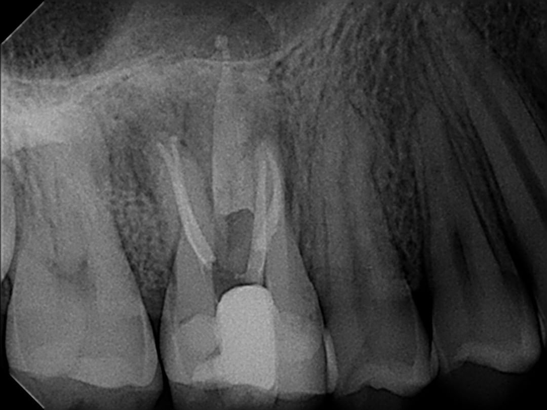
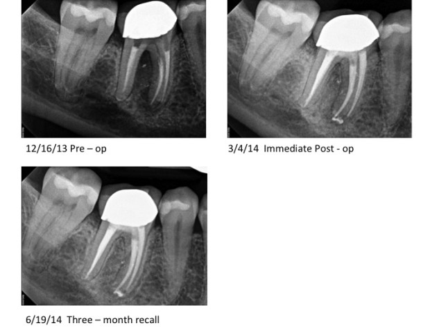
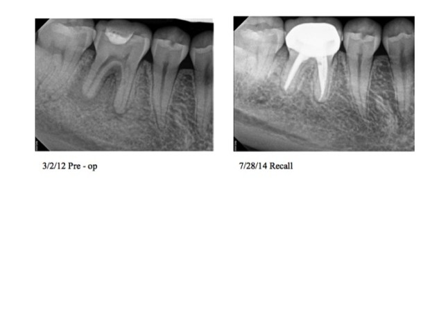
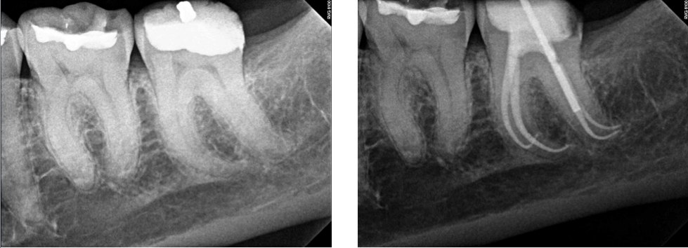
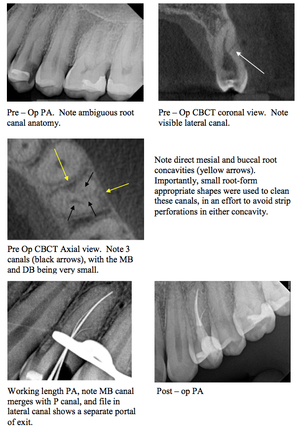

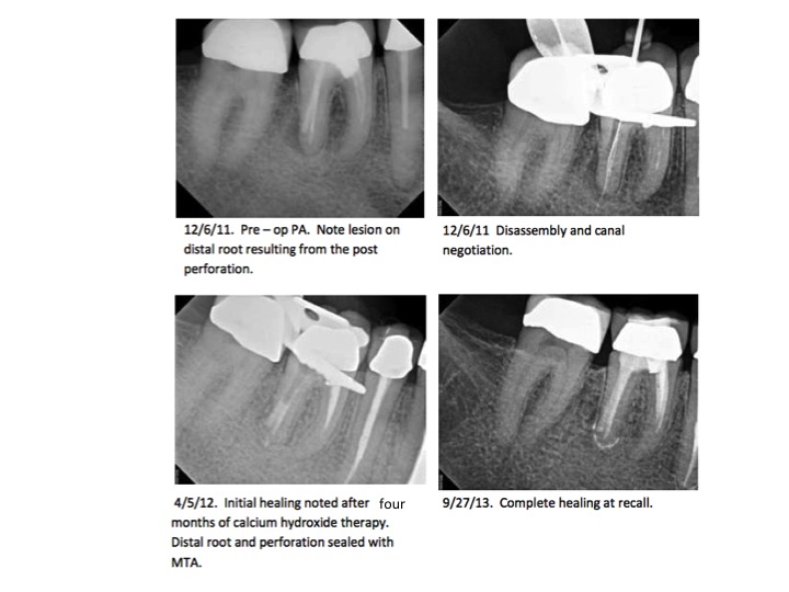
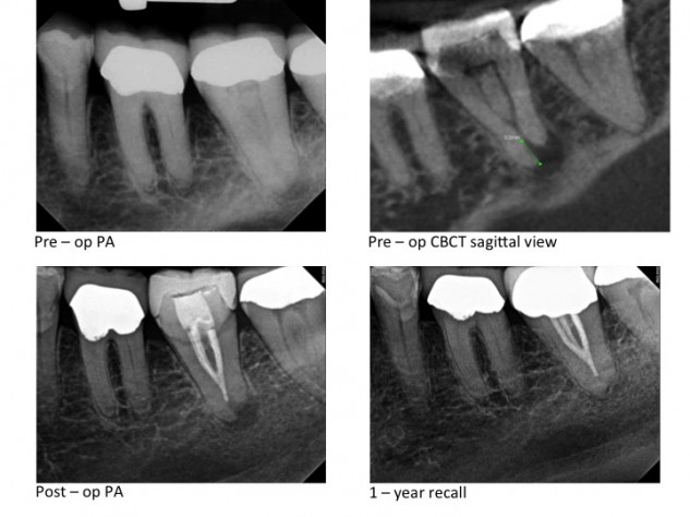 Note how in the case above, the pre-op PA does not indicate where the minor constriction is. The CBCT view shows that the minor constriction is 3.2 mm coronal to the radiographic apex. The case was filled to the minor constriction, and excellent healing is evident at the 1-year recall.
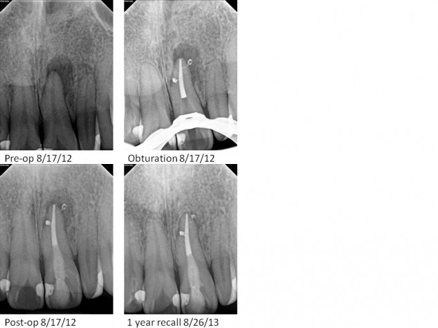
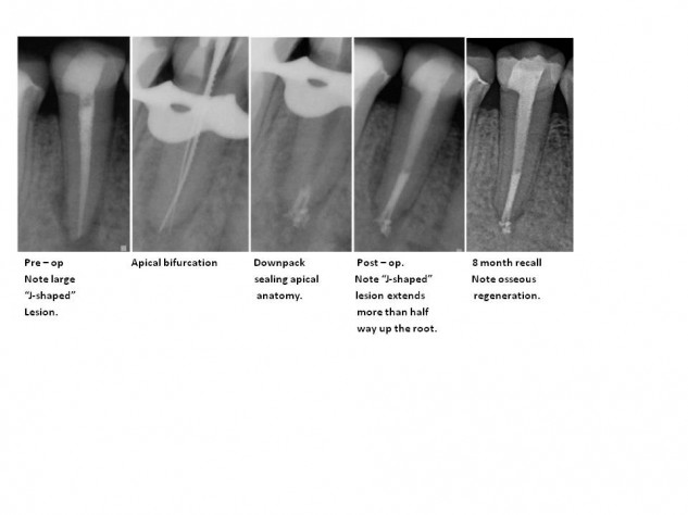
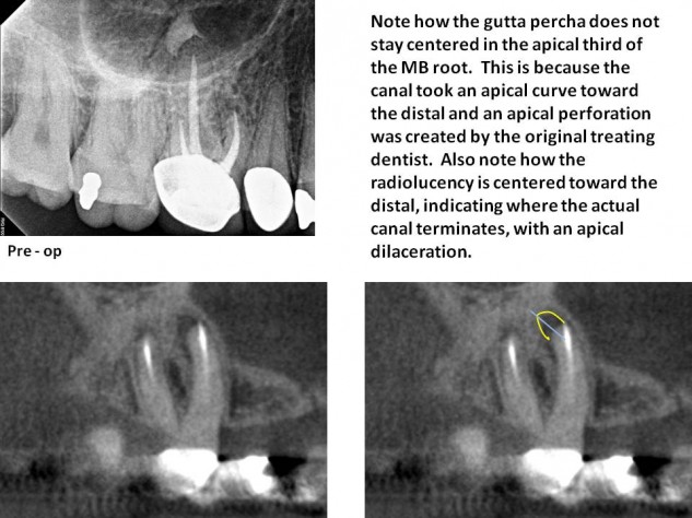
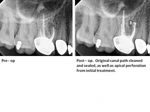
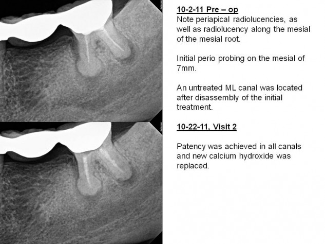
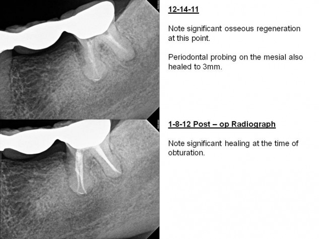
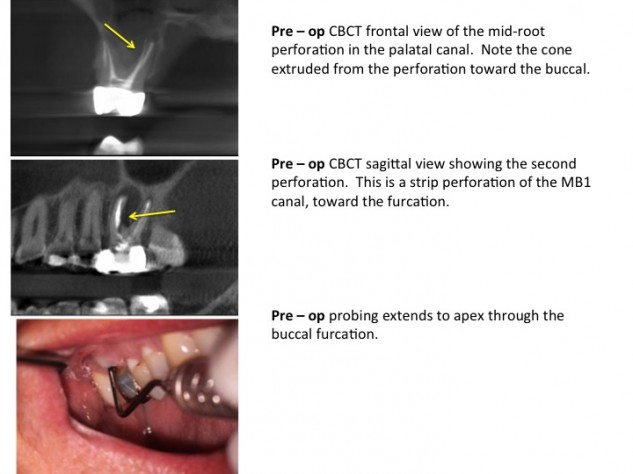
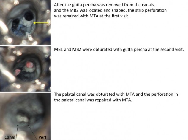
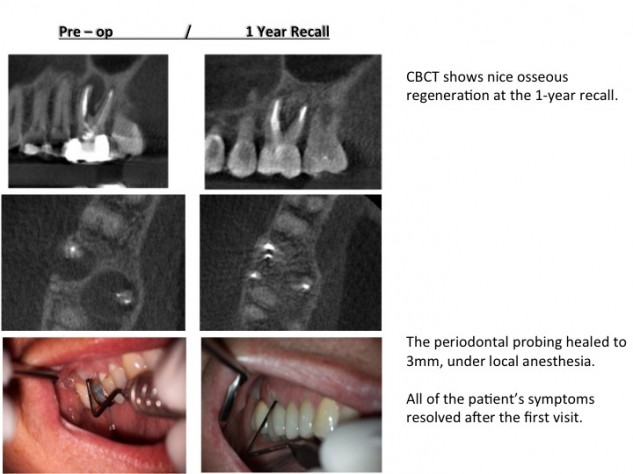
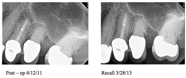 Although the literature may show that there is no difference in treating cases in one vs. two visits, I almost always treat cases with symptomatic apical periodontitis in multiple visits. The main reason is for predictability: I can make sure that we are able to resolve the symptoms prior to finishing the case. A second reason is that we can use calcium hydroxide to help disinfect the root canal system. In the last few years, technological advancements in irrigation have been shown to increase the efficacy of irrigants at disinfecting canals. For patients who have difficulty tolerating dental treatment, the potential exists to treat these case types in a single visit.
 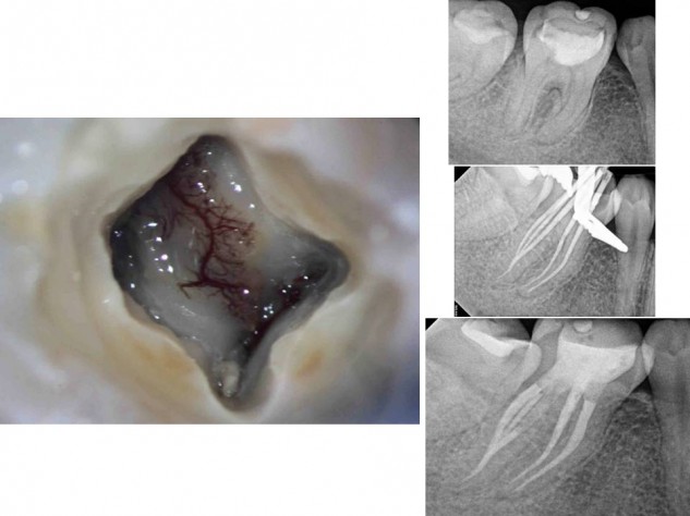 In this case, an indirect pulp cap was done by the patient’s dentist years ago, and recently became symptomatic. Clinical photo of the complex microvasculature in a vital coronal dental pulp. This # 30 had five canals, with the fifth being a mid-distal. Endovac apical negative pressure irrigation is useful even in this case type. Although the main canals were only shaped to a 20-07 and the mid-distal to a 20-04, the microcannula reached beyond where the distal canals merged and allowed for a high flow of irrigants through each canal, to effectively remove vital tissue from the anastamosis. Fiber post bonded in the DL canal.